Four Common Mistakes and How to Improve Experimental Reliability
Cell counting is one of the most essential tasks in cell biology. Whether you’re seeding a 96-well plate, preparing cells for transfection, or monitoring cell proliferation, the accuracy of cell counting has a direct impact on the success of your experiments. Yet, despite its routine nature, many researchers unknowingly repeat avoidable mistakes that can compromise the reliability and reproducibility of their data.
In this article, we highlight four common cell counting mistakes and offer practical solutions to help you achieve consistent, reproducible results.
One of the most frequent errors in cell counting is not mixing the cell suspension properly before loading it. Cells tend to settle quickly at the bottom of tubes or wells, especially if left at room temperature. This results in uneven distribution and highly variable counts from one aliquot to another.
• If your sample from a non-homogeneous mixture, your cell counting will be inconsistent and irreproducible – potentially affecting downstream applications like cell seeding or viability assays.
• Thoroughly mix your cell suspension by pipetting or using a vortex mixer before each cell count
• If using viability dyes like trypan blue, mix again just before loading into the cell counting chamber.
Another common error is incorrect dilution factor calculations after adding dyes like trypan blue, which can lead to significantly inaccurate cell counting results.
Because dilution is defined as the ratio between the total volume and the original cell volume. Since you’re mixing 10 μL of cell suspension with 10 μL of trypan blue, the final volume becomes 20 μL, and the original cell volume is still 10 μL. That makes it a 1:2 dilution, and the dilution factor is calculated accordingly:

To calculate cell concentration, most researchers use the formula:

A hemocytometer’s chamber – typically used for mammalian cell counting – has a standardized depth of 0.1 mm, and each large square has an area of 1 mm².
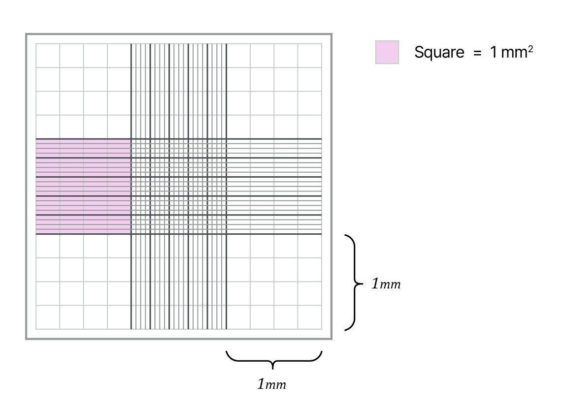
This gives a volume of:

Therefore, to express the number of cells counted per square as a concentration in cells per mL, we multiply by 10⁴
• Always verify both the total volume and the volume of the original cell sample when calculating dilution factors.
• Avoid switching between dilution ratios (e.g., 1:2 → 1:4 → 1:3) across experiments, as this increases error risk. Instead, define and consistently apply a standardized formula and dilution approach in every cell counting step.
• If you’re using an automated cell counter like LUNA-III™ or LUNA-FX7™, standard dilution factors are automatically applied based on the selected protocol, and the device calculates and stores results automatically – helping minimize user error and ensure traceability.
Cell clumps or debris in your sample can lead to serious counting errors. You may undercount if clumps are recognized as single cells, or overcount if debris is mistaken for viable cells.
• Clumps may be mistaken for large single cells
• Debris might be falsely included as viable cells
• Gently resuspend clumped cells through pipetting or mild trypsinization.
• Filter your sample using a 40 µm mesh if necessary.
• When using an automated cell counter (like LUNA-III™ or LUNA-FX7™), you can improve accuracy by adjusting parameters such as size gating, noise reduction, detection sensitivity, and threshold settings to help exclude debris, clumps, and artifacts from your cell counting.
• Especially, LUNA-FX7™ offers advanced parameters – such as declustering controls in fluorescence advanced mode – which help improve the detection of true single cells.
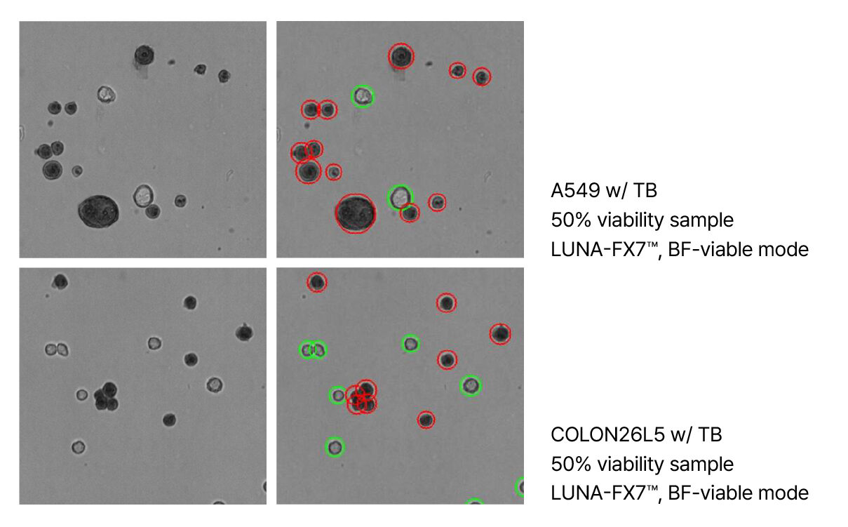
A549(top) and COLON26L5(bottom) cells stained with Trypan blue, imaged using the Brightfield Viable mode of the LUNA-FX7™.
Trypan blue exclusion is widely used for viability analysis, but the timing of analysis is crucial. Over time, live cells can begin to take up the dye, leading to falsely reduced viability readings.
• Perform cell counting within 1–2 minutes after mixing with trypan blue.
• Avoid leaving the mixture sitting in the counting chamber for extended periods.
• Better yet, use more stable viability dyes – such as fluorescence-based reagents (e.g., AO/PI, FDA/PI) or brightfield-compatible alternative like Erythrosine B(EB) – which offer more consistent and less subjective results over time.
▶ Read more: How to choose the right viability stain for automated cell counting
LUNA-FX7™ supports both brightfield and dual-fluorescence cell counting, allowing precise timing, reduced user variability, and improved reproducibility.
The four key mistakes discussed so far – inadequate mixing, dilution factor errors, miscounting clumps or debris, and delayed counting after staining – may seem minor on their own. However, when repeated, these small deviations can seriously undermine the accuracy and reproducibility of your experimental data.
This issue becomes even more critical in shared lab environments, where multiple researchers work with the same samples or data. When cell counting methods, dilution ratios, or viability thresholds differ slightly from one user to another – or when teams alternated between manual and automated counters – comparability across experiments becomes unreliable. Over time, such inconsistencies can erode the credibility of long-term studies and datasets.
• Clearly define and document a standardized SOP for cell counting across your lab
• Align all users on consistent practices for dilution, viability criteria, loading volumes, and counting methods
• When using automated counters like LUNA-FX7™, configure and lock in preset modes and analysis parameters to minimize user-dependent variability
Manual cell counting is not only time-consuming, but also vulnerable to human error, fatigue, and subjective interpretation. Even experienced researchers can introduce variability when performing repetitive tasks.
• Automated calculations and error reduction
• High-resolution image-based cell counting
• Standardized workflows across all users
• Reliable viability assessments using brightfield and fluorescence modes
• Easy data export and documentation for traceability
Automation doesn’t just save time – it provides the consistency and reliability required for high-quality research, especially in labs where multiple users need to generate comparable results.
Cell counting may seem routine, but even small mistakes can lead to inaccurate data, wasted reagents, and failed experiments. By avoiding the four common errors outlined in this article and committing to consistent, validated practices, your lab can ensure reliable, reproducible results—every time.
Whether you’re preparing for high-throughput screening, cryopreservation, or basic culture work, accurate cell quantification is the foundation of every experiment. In today’s research environment, automation is no longer a luxury – it’s a necessity for labs focused on precision, consistency, and efficiency.
Explore the LUNA™ series to count smarter, not harder – with precision, and confidence.
Q1. Why are cell counting results inconsistent even when using the same sample or a hemocytometer?
A. Inconsistencies often result from inadequate sample mixing, miscalculated dilution factors, or misidentification of clumped cells or debris as viable single cells. These issues can occur even with the same sample, especially if aliquots are taken at different times or without thorough mixing beforehand.
Manual counting with a hemocytometer also introduces user-to-user variation based on pipetting skill, visual judgement, and interpretation criteria (e.g., viability). Even within a single lab, inconsistent practices between researchers can significantly affect reproducibility.
To minimize these variations:
• Always mix your cell suspension thoroughly before each count
• Apply a consistent dilution and counting formula across users
• Standardize procedures through a shared SOP
• Consider using automated counters, which eliminate user bias by applying fixed analysis parameters and storing results for traceability
Q2. What should I do if cells are clumping during counting?
A. Resuspend thoroughly by pipetting, use enzymes like trypsin if needed, and filter samples through a 40μm mesh. Automated counters like LUNA-FX7™ offer size gating and declustering to reduce user-dependent variation.
Looking for the Korean version? Click here.
Logos Biosystems provides a diverse portfolio of automated cell counters designed to meet various laboratory requirements.
To learn more, visit www.logosbio.com.


