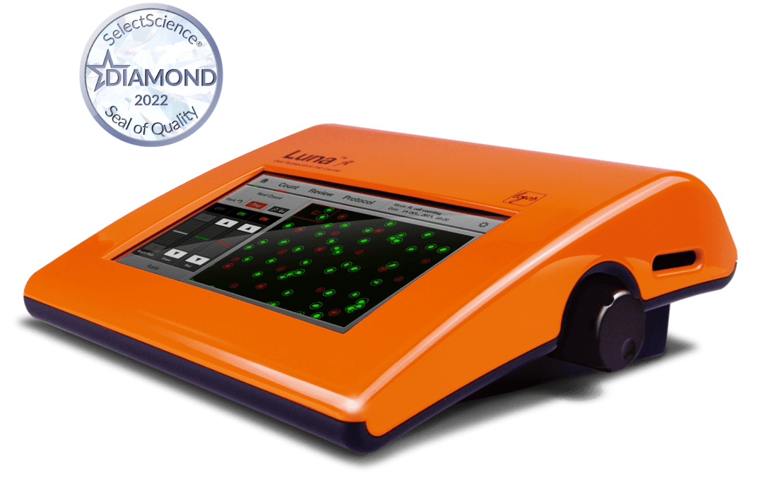
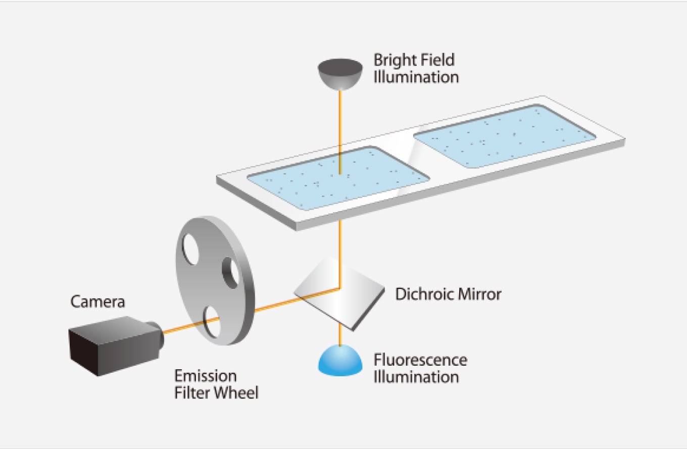
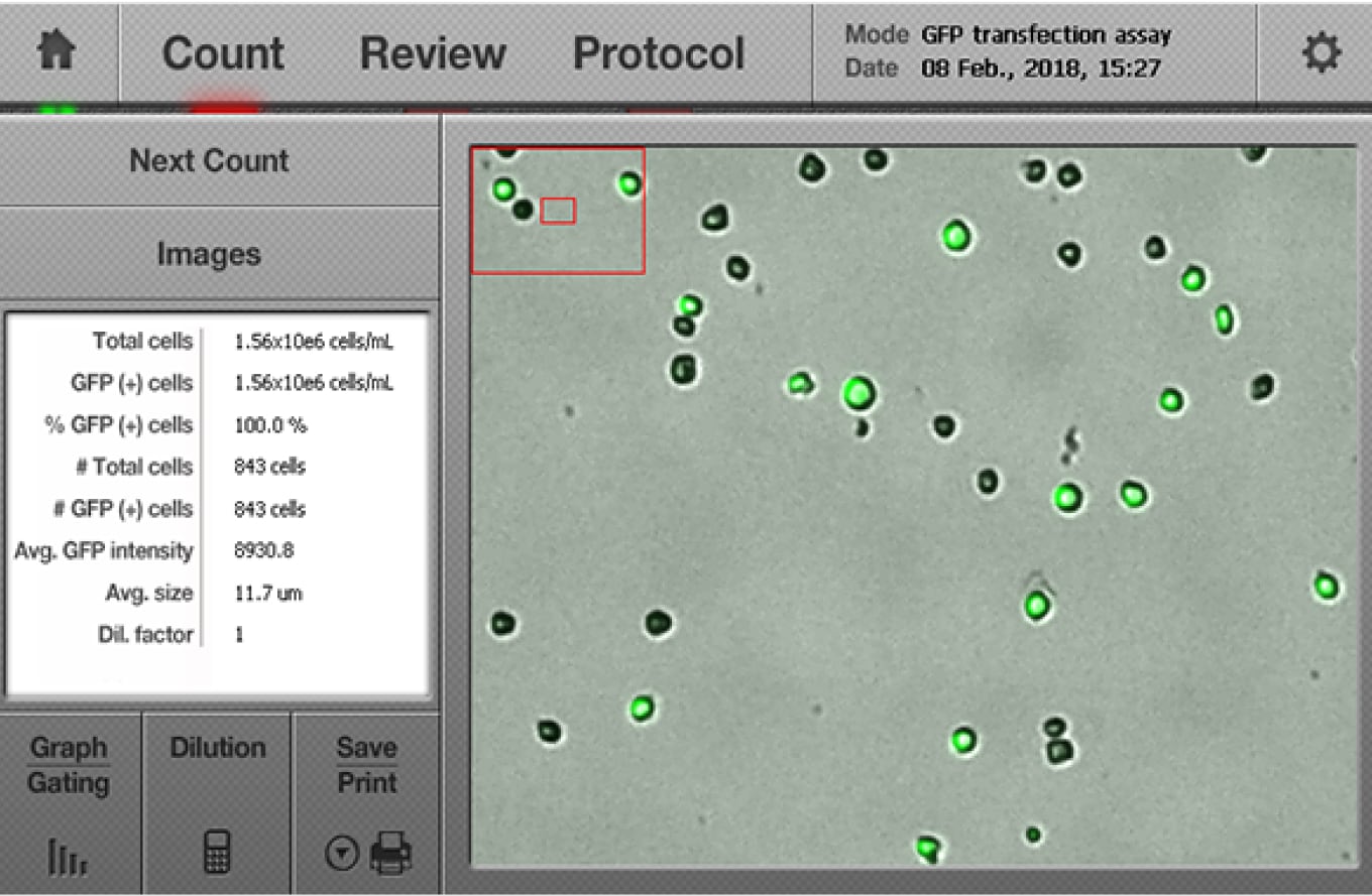
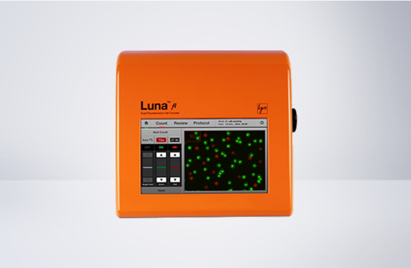
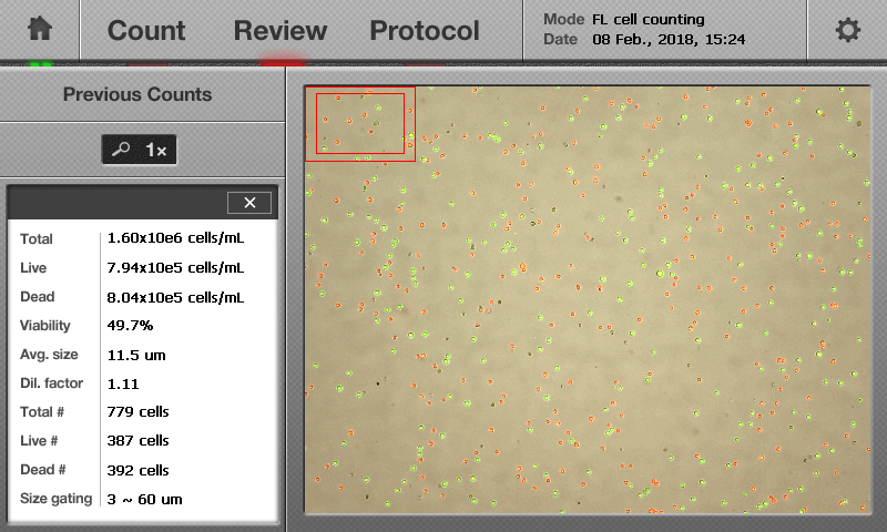
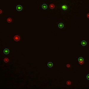
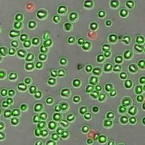
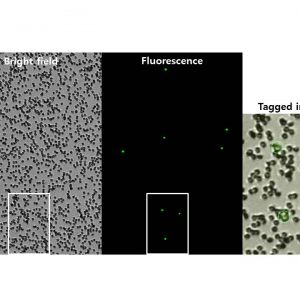
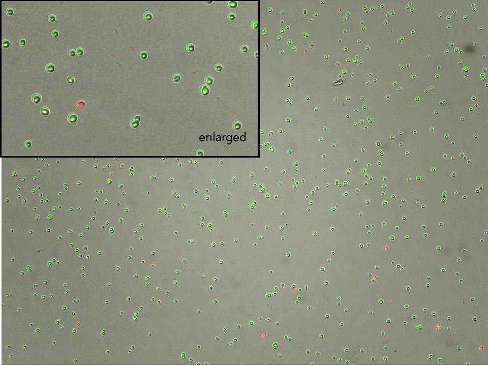
Equipped with dual fluorescence and brightfield optics, the LUNA-FL™ is a powerhouse. The LUNA-FL™ can assess cell counts, viability, and GFP transfection efficiency without being limited by cell type or size.
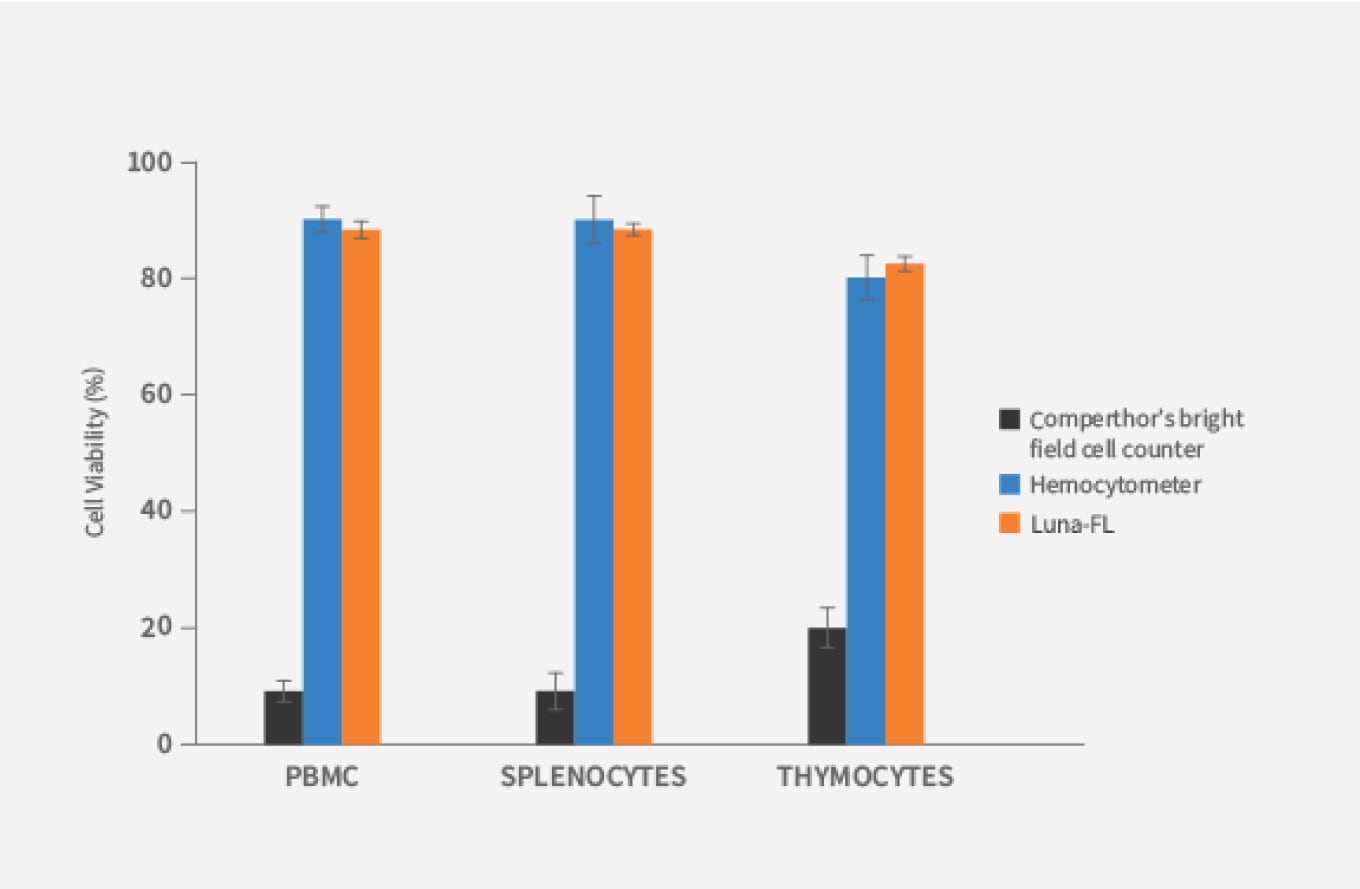
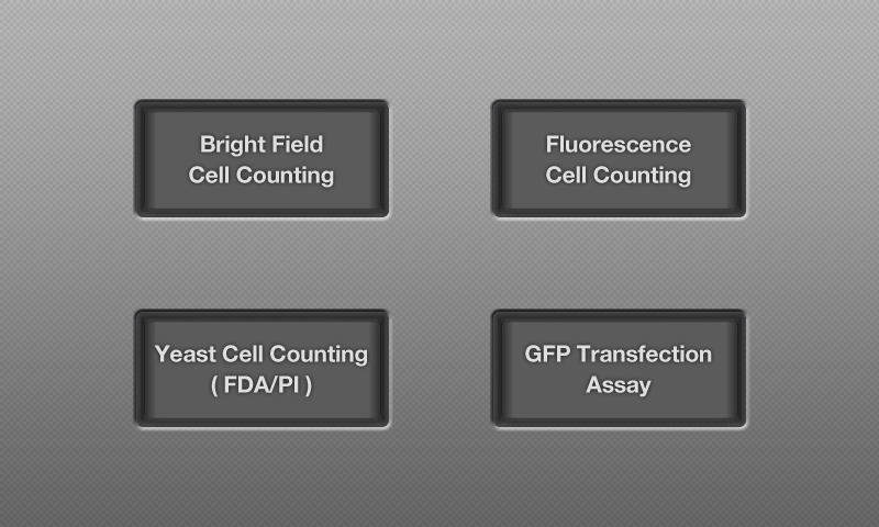
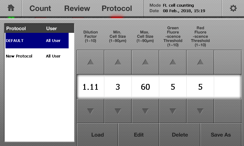
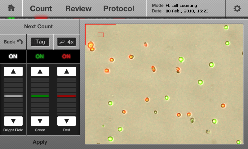
With four counting modes, the LUNA-FL™ is our powerhouse. Equipped with brightfield and dual fluorescence optics that allows the sensitive detection of most cell types excluding bacteria. The LUNA-FL™ can distinguish primary cells such as PBMCs, splenocytes, neutrophils, and stem cells from undesirable debris for accurate cell count and viability results. Brightfield cell counting, fluorescence cell counting, yeast cell counting, and GFP transfection assays are all in the LUNA-FL™ wheelhouse. This versatile little counter sits comfortably in a cell culture hood or on your benchtop to fit seamlessly into your workflow.
Great performance, would recommend for other researchers!
Easy to use, saves us time and offers good quality. The consultant Dr Eberhardt was also professional and patient to teach us before use.
University Heidelberg
Great instrument, I like using it. Really helpful.
The device is easy to use, I mainly use it before cell seeding or to check for my culture's viability and survival. The results are reproducible and the device provides fast results. The accuracy seems to be great as well.
BGU
Brain Cancer Cell-derived Exosomes Protect Scopolamine-Induced Death of SH-SY5Y Neuron Cells.
2021. Lee M. International Journal of Health Sciences and Research 10.36838/v3i1.8.
The Type and Source of Reactive Oxygen Species Influences the Outcome of Oxidative Stress in Cultured Cells.
2021. Goffart S, Tikkanen P, Michell C, Wilson T, Pohjoismäki JLOLO. Cells 10(5):1075.
Immune Memory in Mild COVID-19 Patients and Unexposed Donors Reveals Persistent T Cell Responses After SARS-CoV-2 Infection.
2021. Ansari A, Arya R, Sachan S, Jha SN, Kalia A, Lall A, Sette A, Grifoni A, Weiskopf D, Coshic P, Sharma A, Gupta N. Frontiers in Immunology 12:636768.
The Small GTPase Arf6 Functions as a Membrane Tether in a Chemically-Defined Reconstitution System.
2021. Fujibayashi K, Mima J. Frontiers in Cell and Developmental Biology 9:628910.
Flow cytometric quantification of apoptotic and proliferating cells applying an improved method for dissociation of spheroids.
2021. Metzger W, Rösch B, Sossong D, Bubel M, Pohlemann T. Cell Biology International 10.1002/cbin.11618.
2022-04-07 | 2.12 MB
2022-04-07 | 2.14 MB
2018-04-16 | 110 KB
2018-04-16 | 284 KB
2018-04-16 | 1.7 MB
2018-04-13 | 906 KB
2018-04-16 | 403 MB
2018-04-16 | 862 KB
2018-04-16 | 476 KB
2018-04-13 | 665.84 KB
2018-04-13 | 720.79 KB
Sample Volume | 10 μl |
Cell Counting Time | 30 sec |
Cell Concentration Range | 5×10⁴ – 1×10⁷ cells/mL |
Cell Size Range | Detectable Range: 1 – 90 μm Optimal Range: 5 – 60 μm |
Excitation wavelength | 470 ± 20 nm |
Emission wavelength | 530 ± 25 nm, 600 nm (LP) |
Light Source | LED |
Image Resolution | 5 MP |
LCD Display | 800 x 480 pixels |
Dimensions (W×D×H) | 22 × 21 × 9 cm (8.6 × 8.3 × 3.5 in) |
Weight | 1.8 kg (4 lb) *without the AC adapter and power cord |