Tissue clearing techniques have allowed biologists to acquire high-resolution volumetric images without the need to reduce samples to thin serial sections. One major limitation to some techniques is preserving the signal from endogenous fluorescent proteins (FPs). The required dehydration step of solvent-based tissue clearing methods removes water molecules from tissues, which are required to preserve FP signal. Although recent solvent-based techniques 3DISCO and iDISCO have attempted to address this issue, these methods still can only maintain FP emission for a few days. Although other well-known techniques such as CUBIC, Scale, and PACT preserve FP emission, they are a significant time investment and are limited to small tissue samples. The SWITCH technique utilizes strong fixatives and high temperatures, resulting in a loss of FP signal. In principle, the CLARITY method is the most compatible for FP imaging, but having to construct one’s own electrophoresis chamber with a makeshift cooling system to prevent overheating is a challenge to most.
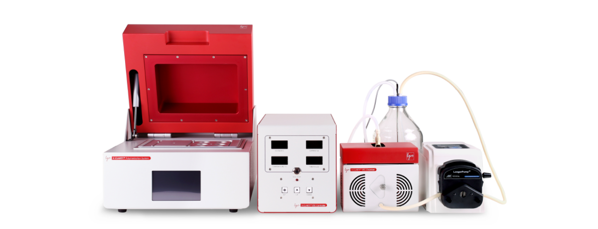
The X-CLARITY™ systems and reagents for tissue clearing are based on the CLARITY principle and have been developed to standardize, simplify, and accelerate each step of the tissue clearing process. One of the components of the X-CLARITY™ Tissue Clearing System is the electrophoretic tissue clearing (ETC) chamber with platinum-plated electrodes and built-in cooling system for efficient tissue clearing. A whole mouse brain takes just 6 hours to clear and endogenous FP signals are preserved. In this report, we present a protocol for sample processing for volumetric imaging using the X-CLARITY™ systems and reagents for tissue clearing with a focus on the preservation of FP signals.
All animal experimental procedures were conducted in accordance with KBRI IACUC guidelines. Adult Thy1-YFP mice (20 weeks old) were anaesthetized with Avertin (250 mg/kg, Sigma) and perfused transcardially with 30 mL PBS, followed by 20 mL fresh 4% PFA (10 mL/min). Brains were extracted and incubated in 4% PFA for 24 hours at 4 °C. Brains were then washed and stored in PBS for 24 hours at 4 °C.
Technical Tips:
– Post-fixation is an important step in the X-CLARITY™ protocol. A whole mouse brain should be post-fixed for at least 24 hours. Excessive fixation can lead to increased clearing time. – Post-fixed tissues should be washed for at least 24 hours as residual PFA can react with acrylamide during hydrogel polymerization and lead to increased clearing time.
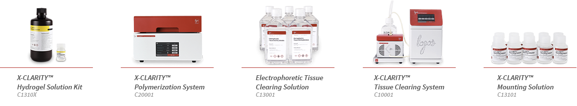
A hydrogel solution was prepared using the X-CLARITY™ Hydrogel Solution Kit (Logos Biosystems, C1310X). Brains were incubated in hydrogel solution (5 mL/brain) for 24 hours at 4°C. Brains in solution were placed in the X-CLARITY™ Polymerization System (Logos Biosystems, C20001) for 2 hours at 37°C and -90 kPa to initiate polymerization.
Technical Tips:
The main difference of the original CLARITY and X-CLARITY™ chemistry is the use of bis-acrylamide and PFA in hydrogel solution. The use of bis-acrylamide and PFA leads to higher cross-liking density, but this also leads to increased clearing time. – Since bis-acrylamide is not used in X-CLARITY™ chemistry, gelation is not observed after hydrogel polymerization. If the liquid hydrogel solution has become a sticky, more viscous solution, acrylamide polymerization has been successful.
The hydrogel-embedded brains were rinsed with Electrophoretic Tissue Clearing Solution (Logos Biosystems, C13001) and then placed in the ETC Chamber of the X-CLARITY™ Tissue Clearing System (Logos Biosystems, C10001). Electrophoretic Tissue Clearing Solution was circulated through the chamber and 0.7 A was applied across the brains for 6-12 hours at 35°C. After clearing, brains were washed with PBS overnight at room temperature to remove residual SDS.
Technical Tips:
– Endogenous FP signals can degrade at higher temperatures. To prevent excessive Joule heating, optimize the electric current applied during electrophoresis. Use lower currents to minimize high temperature increases. This may also affect clearing time.
Clarified brains were cut into 1 mm slices or hemispheres for immunostaining. Brain samples were incubated in anti-Collagen IV (1:100, Abcam) in 6% BSA, 0.2% Triton X-100, 0.01% sodium azide in 0.1 M PBS for 24 hours at 37°C with gentle shaking. After 24 hours, samples were washed with PBST (0.2% Triton X-100, 0.01% sodium azide in 1X PBS) overnight at 37°C. Samples were then incubated with donkey anti-rabbit Cy3 Fab fragment (1:250, Jackson ImmunoResearch) and TO-PRO-3 Iodide (1:1000, ThermoFisher Scientific) in 6% BSA, 0.2% Triton X-100, 0.01% sodium azide in 0.1 M PBS for 24 hours at 37°C with gentle shaking. Samples were washed with PBST overnight at 37°C.
Stained samples were rinsed with distilled water for 5 minutes and immersed in X-CLARITY™ Mounting Solution (Logos Biosystems, C13101) for 1 hour at room temperature with gentle shaking. The solution was replaced and samples were incubated for another 1-2 hours. For confocal imaging, brain slices were placed in a 35 mm glass-bottomed dish (SPL Life Sciences) with fresh mounting solution and imaged with a LSM 710 (Carl Zeiss) using the EC Plan-Neofluar 10x/0.3 objective. ZEN software (Carl Zeiss) was used to process the images. For light sheet imaging, brain hemispheres were placed in an imaging chamber with fresh mounting solution and imaged with a Lightsheet Z.1 (Carl Zeiss) using a EC Plan-Neofluar 5x/0.16 objective lens. ZEN (Carl Zeiss) software was used to process confocal images. Amira 3D software (FEI) was used to process light sheet images and render a 3D video.
Technical Tips:
– Fluorescence imaging should be done immediately after RI matching to minimize the loss of fluorescence signal. Stained samples can be stored in the mounting solution for up to 3 days without significant signal loss, but for long term storage, place samples in PBS at 4°C. – For more information, read “The X-CLARITY™ Mounting Solution: an improved RI matching solution for tissues cleared by the X-CLARITY™ Tissue Clearing System”.
Endogenous Thy1-YFP was stable and well-preserved upon clearing with the X-CLARITY™ systems and reagents. Anti-Collagen IV antibody and TO-PRO-3 Iodide penetrated the sample efficiently. Light sheet images were rendered into a high resolution 3D video and can be viewed here: A journey through a Thy1-YFP mouse brain.
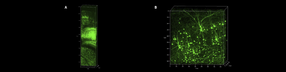
Long-term preservation of the Thy1-YFP signal after clearing with the X-CLARITY™ systems and reagents. (A) Thy1-YFP signal immediately after clearing. (B) Thy1-YFP signal one month after clearing.
In this study, we analyzed the effects of plasmid concentration on transfection efficiency using the CELENA® X High Content Imaging System. Images of the adherent HeLa cells in a multi-well plate were captured automatically and batch processed to identify objects, measure fluorescence intensity, apply a fluorescence intensity threshold, and even label images to help visualize data. As shown in Figure 1 and 2, transfection efficiency increased with plasmid concentration.Using the CELENA® X for fluorescence cell imaging and to quantitatively analyze multiple cellular features from each image automatically gives researchers a simple way to monitor transfection-based cell assays. The same analysis pipeline can easily be reused to verify or compare results from subsequent experiments.


