Key features
| Generating high-resolution panoramic images using the CELENA® X High Content Imaging System. |
Introduction
Capturing an image (or images) gives researchers a visual representation that serves to validate data. Often, a limited field of view (FOV) makes it difficult to capture a clear and concise image. To overcome this limitation, many companies have developed image stitching algorithms. Image stitching is the assembly of multiple images that are ‘stitched’ together to form a high-resolution panoramic image. However, many stitching algorithms often have distortion errors which result in poor image stitching quality. The CELENA® X Cell Analyzer software uses a proprietary image stitching algorithm that produces seamless stitching results. In this study, we demonstrate the seamless image stitching ability of the CELENA® X Cell Analyzer software to form a high-resolution panoramic image.
Materials and Methods
All Hematoxylin and Eosin (H&E) stained samples and organoid samples were obtained from Seoul Asan Medical Center. Organoid samples were stained with YAP1 antibody (Santa Cruz, sc-101199), Alexa Fluor™ 594 Phalloidin (Life Technologies, A12381), and Hoechst 33342 (Life Technologies, H3570). H1975 Cells were plated on a 96-well plate and stained with MitoTracker™ Red (Life Technologies, M7512), Acridine Orange (AO; Logos Biosystems, F23002) and Hoechst 33342 (Life Technologies, H3570). The samples were imaged on the CELENA® X High Content Imaging System (Logos Biosystems, CX30000). The CELENA® X Cell Analyzer software was used to complete the image stitching by using the image stitching pipeline. To compare image stitching performance, Company A’s automated system and analysis software was also used following the same method and materials.
Imaging
Cells and organoid samples were imaged using a 10X LWD objective, and H&E samples were imaged using a 20X LWD objective. A DAPI filter cube (Ex395/25, Em460/50), an EGFP filter cube (Ex470/30, Em530/50), and a RFP filter cube (EX530/40, Em605/55) were used for multi-channel imaging.
Improved imaging stitching algorithm
Image stitching algorithm works to pair images based on the similarity of the overall appearance between images. In general, image stitching is performed following the order of how the images are saved. Unfortunately, this strategy can cause unwanted visual artifacts to appear along the image boundaries. This is especially true if there are no distinct characteristics present. This issue is a common problem when using image stitching algorithms (Figure 1A). We resolved this issue by assembling distinct images first instead of following the order in which the images were saved. In turn, the CELENA® X Cell Analyzer software provided seamless image stitching results even with less distinguishable images (obtained from the upper part of the stitched image). (Figure 1B).
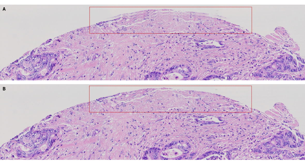
Figure 1. (A) The final result of the stitched image shows it was misaligned when using the original image stitching algorithm. (B) The final result of the stitched image shows a seamless image using the advanced proprietary algorithm. (1500 x 375 pixels)
Superior quality of image stitching
Image stitching performance was compared between the CELENA® X Cell Analyzer software and company A’s software. The comparison was made by running the image stitching algorithm with images obtained from the same sample. The stitched image from company A’s software had distortion and misalignment errors which made it difficult to accurately understand the overall morphology of each sample. (Figure 2A). Whereas the CELENA® X Cell Analyzer software successfully produced a seamless image with no visible errors (Figure 2B).
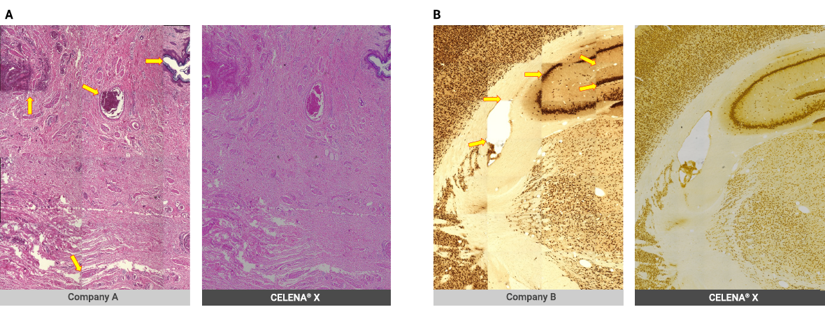
Figure 2. (A) Image stitching results using H&E slides: Company A’s image results show misalignment and distortion issues versus the result image using the CELENA® X Cell Analyzer Software and the seamless image results (301 x 423 pixels). (B) Image stitching results using mouse brain sample: Company A’s image results show misalignment and distortion issues versus the result image using the CELENA® X Cell Analyzer Software and the seamless image results (471 x 662 pixels).
Whole well image stitching
The CELENA® X High Content Imaging System is designed to run in vitro scientific experiments. The complete regions of interest on multi-well plates and dishes can be selected and imaged by the CELENA® X Cell Analyzer software. With this advantage, the whole-well image stitching was conducted using multi-channel images to show superior image stitching performance (Figure 3).
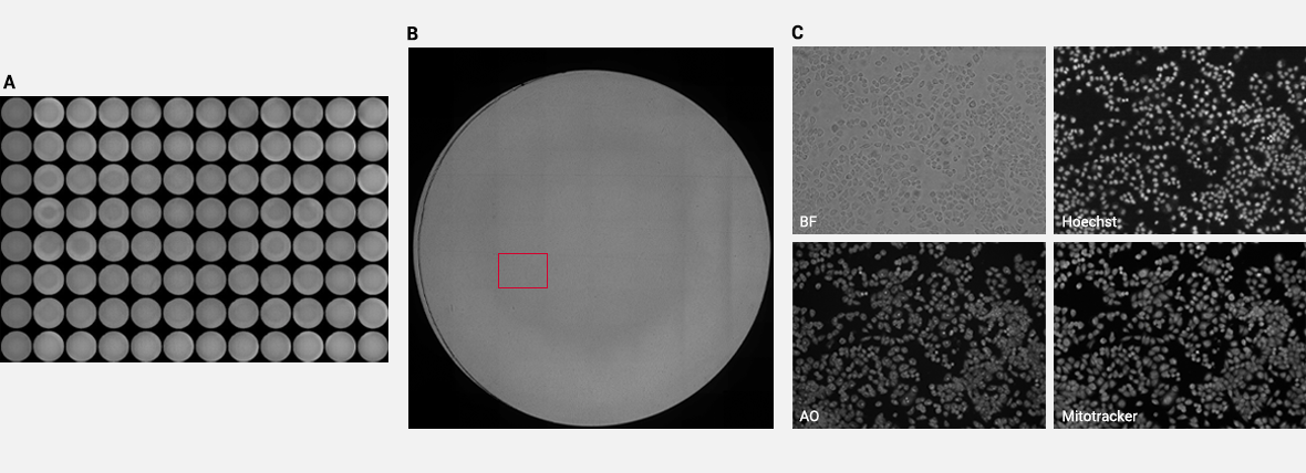
Figure 3. (A) The whole 96-well plate image stitching result. (B) The image stitching result from one of 96 wells using BF images (11336 x 11788 pixels). (C) Multi-channel images obtained from the area marked with the red square.
Image stitching on organoid models
Organoid culture is a three-dimensional cell culture technique mimicking the physiological conditions like that of organs. Thanks to this property, experiments using organoid culture provide results similar to in vivo experiments. Image stitching is a great way of studying organoid samples which can be prepared by sectioning and using immunofluorescent staining to visualize molecular markers. Multi-channel imaging was conducted on organoid samples and image stitching was performed using the CELENA® X High Content Imaging System (Figure 4).
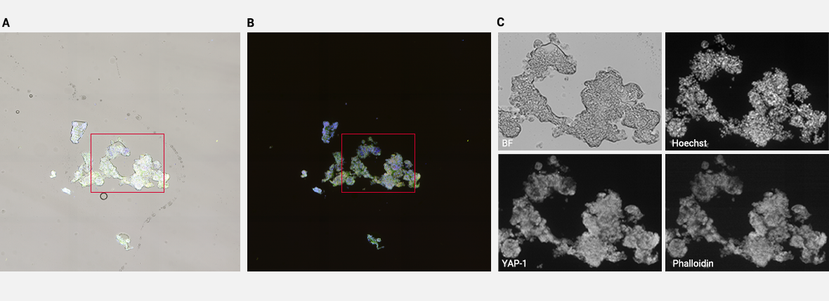
Figure 4. The combined image stitching results using all channels with (A) and without (B) BF channel (4464 x 4428 pixels). (C) Multi-channel images obtained from the area marked with the red square.
Conclusion
Here, we have shown the advanced quality of the CELENA® X Cell Analyzer software’s proprietary algorithm for image stitching compared to the image stitching algorithms widely used by other companies. The comparison between result images from Company A’s software and the CELENA® X Cell Analyzer software clearly showed precise, clear image stitching performance when using CELENA® X Cell Analyzer software. Moreover, the superior image stitching quality was confirmed through in vitro imaging on a whole 96-well plate as well as using organoid samples. In conclusion, the CELENA® X High Content Imaging System with its proprietary image stitching used with the CELENA® X Cell Analyzer software, would be advantageous for researchers and scientists for capturing optimal panoramic images.


