Cell counting is an essential step in routine cell maintenance and for obtaining accurate and consistent experimental results. Among its many features, the CELENA® S Digital Imaging System has an automated cell counting mode. Users can select cell detection parameters to count different types of cells and the CELENA® S does the rest, adjusting focus and light for optimal cell detection. An image of the cells is captured, analyzed, and labeled to automatically distinguish live and dead cells. Cell count and viability results appear next to the image and can be exported easily via USB. In this study, we compared the cell counting performance of the CELENA® S to an automated cell counter and manual cell counting with a hemocytometer.
Sample preparation
Series of HL-60 and HEK-293 cell suspensions with different viabilities were prepared by mixing live and dead cells. Live cells were prepared from exponentially growing cells. Dead cells were prepared by incubating an appropriate number of cells at 70°C for 30 minutes. Cells were mixed with an equal volume of 0.4% trypan blue prior to counting.
Counting Methods
A CELENA® S Digital Imaging System, LUNA-II™ Automated Cell Counter, and a Marienfeld glass hemocytometer with Neubauer counting grids were used to determine cell concentration and viability. When counting with the CELENA® S and LUNA-II™, cells were loaded into a compatible disposable slide and counted with the DEFAULT protocols of each with Autofocused Counting activated. When counting with the hemocytometer, the cells within the two corner squares of the Neubauer counting grids were counted. All experiments were performed in triplicate.
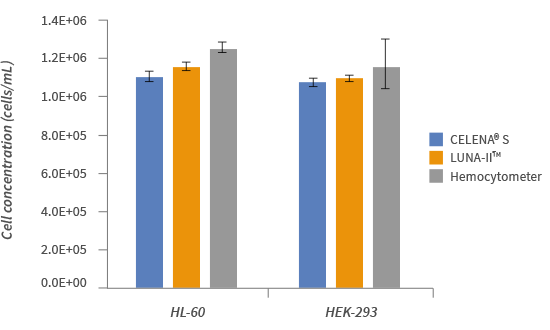
Figure 1. Comparison of three cell counting methods.
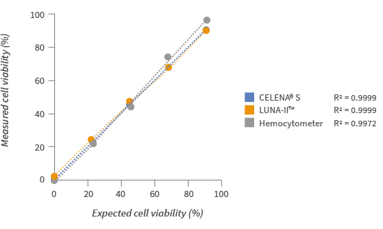
Figure 2. BLinearity and accuracy of cell viability analysis using samples with different expected viabilities.
To determine the cell counting accuracy of the CELENA® S, cell samples with a total concentration of approximately 1 x 10⁶ cells/mL were counted with the CELENA® S, the LUNA-II™, and a hemocytometer. All three methods produced relatively similar results (Fig. 1). Hemocytometer results were slightly higher on average but more variable from count to count (Fig. 1). When assessing the accuracy of cell viability analysis, CELENA® S results showed a high degree of linearity (R2 > 0.999) with the expected viabilities (Fig. 2).
The CELENA® S is a small and powerful digital imaging system that can be used for multiple applications, such as capturing and analyzing multicolor fluorescence images, live cell imaging, z-stacking, and automated cell counting.
The automated cell counting feature is a quick, accurate, and reliable way to count stained cells.
Accurate – cell count and viability measurements
Consistent – definite parameters (such as cell size and shape) used to detect cells to eliminate variability
Convenient – captured images and data easily exported via USB
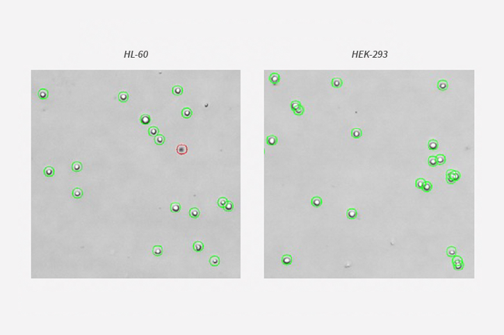
Figure 3. Cell images captured and analyzed with the automated cell counting feature of the CELENA® S. Live and dead cells are labeled with green and red circles, respectively.
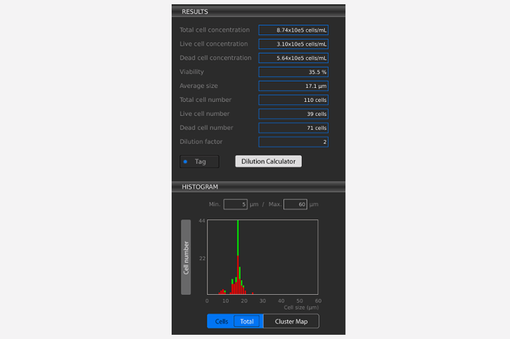
Figure 4. Cell count results on the CELENA® S .


