Practical Tips to Improve Transfection Efficiency
: From Cell Health and Toxicity Control to Quantitative Evaluation
In cell-based life science research, transfection is one of the most fundamental yet critical techniques.
Successfully delivering a gene of interest into cells to induce protein expression, or suppressing the expression of a specific gene through knockdown or knockout approaches, directly determines the reliability of experimental outcomes.
However, in day-to-day laboratory practice, researchers frequently encounter the following challenges:
Low transfection efficiency
Excessive cytotoxicity
Poor reproducibility between experiments
A significant portion of these issues arises not from the intrinsic performance of the transfection reagent itself, but from basic experimental conditions that are under the researcher’s control.
To better understand these common challenges associated with transfection experiments, we will first briefly review the fundamental concepts, and then highlight key practical points that can be readily applied in the laboratory to improve experimental outcomes.
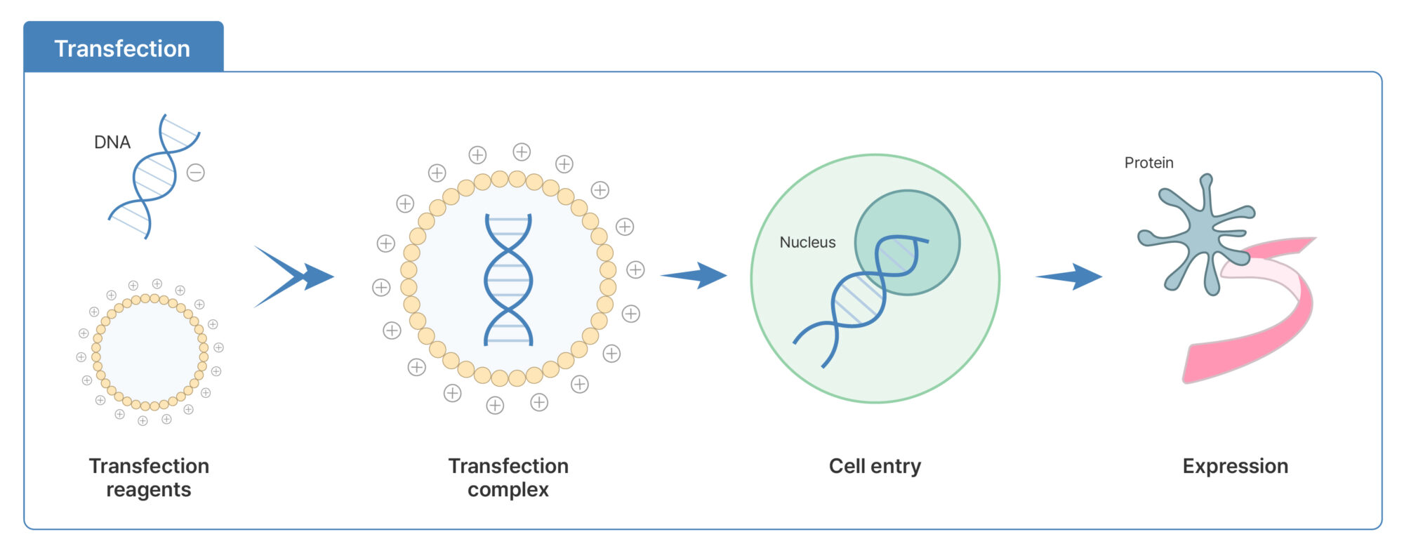
Figure 1. Basic principle of transfection DNA forms a complex with a transfection reagent, enters the cell, and subsequently leads to gene expression in the nucleus.
What is Transfection?
Transfection is a technique used to artificially introduce nucleic acids, such as DNA or RNA, into eukaryotic cells using non-viral methods in order to induce gene expression. After forming a complex with a transfection reagent, the nucleic acid enters the cell and, depending on the cell cycle and delivery mechanism, reaches the nucleus where gene expression occurs (see Figure 1).
Cationic reagents (+) electrostatically interact with negatively charged DNA (–) to form complexes, which are able to cross the cell membrane and ultimately drive protein expression within the nucleus.
The main purposes of transfection include:
Functional analysis of specific genes
Protein expression and reporter assays
Gene silencing using siRNA or miRNA
■ Representative Transfection Methods
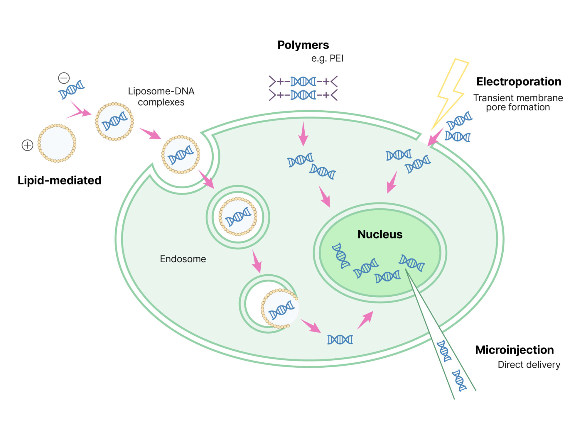
Chemical methods These methods use cationic lipid- or polymer-based reagents to form DNA/RNA–reagent complexes that facilitate passage across the cell membrane.
Physical methods Techniques such as electroporation or microinjection create transient pores in the cell membrane through electrical pulses or direct mechanical injection.
Among these approaches, lipid- or polymer-based transfection methods are the most widely used due to their accessibility and broad applicability. Figure 2 compares the mechanisms of action for each method. Lipid-mediated transfection delivers DNA via endocytic pathways, whereas electroporation introduces nucleic acids through transient membrane pores generated by electrical pulses. In contrast, microinjection delivers nucleic acids directly into the nucleus using a fine needle.
Figure 2. Comparison of representative non-viral transfection methods A conceptual illustration of the mechanisms underlying chemical (lipid- and polymer-mediated) and physical (electroporation and microinjection) transfection approaches.
■ Comparison of Transfection, Transduction, and Transformation
Although these terms may appear similar in that they all involve the delivery of genetic material into cells, they represent distinct concepts and should be clearly differentiated.
Prokaryotic cells (Bacteria) or plant/fungal cells
Introduction of plasmid or other exogenous DNA
As summarized in the table above, transfection refers to the introduction of nucleic acids into eukaryotic cells using non-viral methods, resulting in either transient or stable gene expression. In contrast, transduction relies on viral vectors (such as lentiviruses or adenoviruses) to achieve stable gene delivery, while transformation is primarily used for genetic manipulation in bacteria, such as the introduction of plasmid DNA.
Among these approaches, transfection is the most suitable method when rapid expression analysis and experimental design flexibility are required.
Why Does Transfection Efficiency Vary?
Many researchers tend to attribute low transfection efficiency solely to the performance of the transfection reagent. However, in practice, the physiological state of the cells and the experimental environment often have a greater impact on efficiency variability.
Transfection efficiency is not determined by a single parameter; rather, it is influenced by a combination of multiple factors, including:
Physiological condition of the cells
Plating density (confluence)
DNA quality and reagent conditions
Management of cytotoxicity
Methods used to evaluate transfection efficiency
If even one of these factors is inconsistent, the reproducibility of experimental results can be significantly compromised.
Five Practical Tips to Maximize Transfection Efficiency
Transfection efficiency is influenced not only by the quality of the reagent or DNA, but also to a large extent by cellular and experimental conditions that are directly controlled by the researcher.
Tip 1. Cell Health and Confluence Cell condition is one of the most critical determinants of transfection efficiency.
Cell health
Cells should be in the logarithmic growth phase (log phase) at the time of transfection. Cells that have undergone excessive passaging (high passage number) often show reduced transfection efficiency and poor experimental reproducibility.
Confluence
For most adherent cell lines, a confluence of approximately 60–80% at the time of transfection generally yields more consistent and reliable results.
Too low: increased cellular stress and susceptibility to cytotoxic effects
Too high: reduced gene expression due to cell cycle arrest or limited cell division
Culture medium condition
Replacing the culture medium with fresh medium immediately prior to transfection can help reduce the accumulation of metabolic byproducts and potential cytotoxic factors released by the cells.
Tip 2. DNA/RNA Quality and Concentration
DNA quality
Plasmid DNA should be purified under endotoxin-free conditions whenever possible. Endotoxins can induce cytotoxic effects, negatively impacting both transfection efficiency and overall cell viability.
Concentration optimization
Increasing the amount of nucleic acid does not necessarily lead to higher transfection efficiency. Excessive amounts of DNA or RNA may instead increase cytotoxicity and compromise overall experimental quality. Gradual optimization based on the manufacturer’s recommended conditions is therefore essential.
Tip 3. Reagent Selection and Complex Formation
Reagent selection
The performance of transfection reagents can vary depending on the cell type and experimental objective. It is generally advisable to begin with reagents that have been broadly validated across multiple cell lines.
Complex formation conditions
DNA and transfection reagents are typically mixed in serum-free medium or buffer to allow efficient complex formation. Serum components may interfere with complex formation and reduce transfection efficiency.
Complexation time
The duration of complex formation is critical; both insufficient and excessive incubation times can negatively affect transfection performance. Maintaining a consistent complexation time across experiments is therefore important for reproducibility.
Tip 4. Toxicity Management
Transfection reagents inherently impose stress on cells.
Replacing the culture medium at an appropriate time point after complex addition (typically 4–6 hours) can help reduce cytotoxicity caused by prolonged exposure to the transfection reagent.
Under highly toxic conditions, gene expression levels may appear high; however, the overall reliability of the experimental results is often compromised.
In transfection experiments, “manageable efficiency” is more important than simply achieving the highest apparent efficiency.
Tip 5. Maintaining Experimental Consistency
Even minor variations in experimental conditions can lead to substantial differences in transfection outcomes, including:
Cell number per well
Culture medium volume
Duration of reagent exposure
To obtain reproducible and reliable results, it is essential to maintain all experimental conditions as consistently as possible across wells and experiments.
Transient vs. Stable Transfection (Basic Overview)
Transient transfection
Introduced genes are expressed only temporarily, making this approach well suited for rapid evaluation of gene expression and short-term experiments.
Stable transfection
The introduced gene is integrated into the host cell genome, and a stable cell line is established through antibiotic selection, enabling long-term and consistent gene expression.
While the generation of stable cell lines is advantageous for long-term studies, it requires significantly more time and extensive optimization of experimental conditions.
Making Transfection a Manageable Process
Transfection efficiency cannot be optimized by adjusting a single variable alone. Effective transfection requires a comprehensive approach that considers cell health and confluence, reagent and nucleic acid conditions, and the methods used to evaluate efficiency.
Rather than simply determining whether GFP expression is “visible” under a microscope, the use of quantitative analysis tools allows transfection outcomes to be objectively compared and validated using measurable data. Instead of memorizing highly specific conditions for each individual cell line, maintaining consistent experimental parameters and evaluating results based on quantitative metrics leads to more stable and reproducible outcomes over time.
Ultimately, the key is to recognize transfection not as a chance-dependent experiment, but as a controllable and manageable process. The five tips introduced in this article—cell health management, DNA quality, reagent conditions, toxicity control, and experimental consistency—are all factors that researchers can directly regulate. When combined with quantitative analysis, these strategies can make transfection experiments far more predictable and reliable.
Completing Transfection Management Through Quantitative Analysis
Quantitative analysis using GFP reporters can be applied in different ways depending on the experimental objective and stage.
Rapid Assessment of Transfection Efficiency: Fluorescent Cell Counting
This approach enables rapid quantification of the percentage of GFP-positive cells within the total cell population. Fluorescent cell counters, such as LUNA-FL™, are particularly useful for quickly comparing multiple transfection conditions and screening for optimal parameters.
→ This method is well suited for the optimization phase, allowing rapid testing of reagent concentration, DNA amount, and incubation time.
In-Depth Analysis: Automated Imaging
Using high-resolution fluorescence imaging systems such as CELENA® X, the extent of transfection under different experimental conditions can be quantitatively compared using image-based analysis following GFP transfection.
Proportion of GFP-positive cells relative to the total cell population
Differences in expression intensity across experimental conditions
Area-based comparison of GFP-positive signals
→ These analyses are useful for objectively comparing and validating differences in transfection efficiency across multiple experimental conditions.
📄 Evaluating transfection efficiency using the CELENA® X
These quantitative analysis approaches help transform transfection experiments from those that merely appear successful into experiments that can be objectively evaluated and explained using data.
Learn More
Interested in exploring more cell counting solutions?
Visit www.logosbio.com to discover a wide range of cell counter solutions tailored to different research goals and analytical workflows.