Keywords: High-quality bacteria imaging, bacteria sample preparation, centrifugation, Brownian motion, z-plane scattering, focus issues, microbial cell visualization, CELENA® X High Content Imaging System
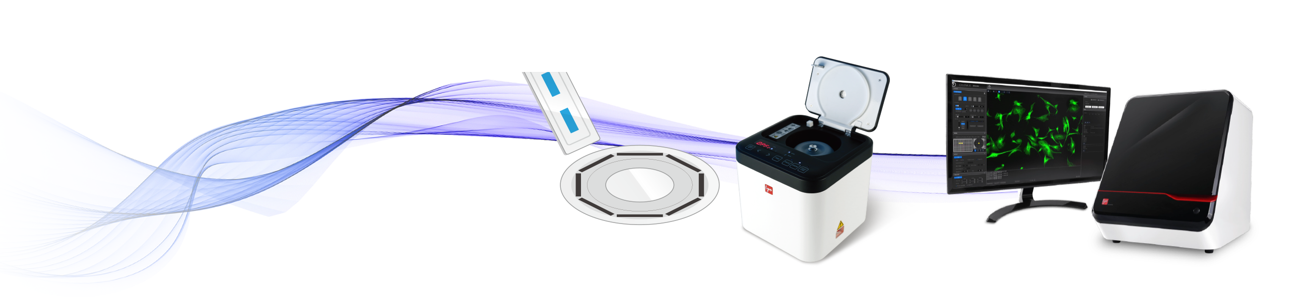
The high-quality imaging of bacteria is a significant challenge due to their minute size and low mass, leading to continuous, erratic movements caused by Brownian motion and scattering across different z-planes. Previous studies have explored various approaches to overcome these issues, including agarose pads or hydrogels to reduce Brownian motion. In addition to mitigating Brownian motion, researchers have investigated conductive coatings on surfaces to induce electrostatic interactions by using materials like poly(L-lysine) to attract bacterial cells. Further investigation has also extended to methods involving negatively charged surfaces and positively charged surfaces to gather all cells into one plane. However, these methods can have issues such as time-consuming sample preparation, high cost and potential focus issues during imaging. A more straightforward method to address focus problems is by using a coverslip on a glass slide, but it has limitations such as Brownian motion and the ability to hold only a restricted sample volume. To address these limitations, we present the new method combining the CELENA® X High Content Imaging System for imaging with the established procedure for QUANTOM Tx™ Microbial Cell Counter to prepare bacterial samples. This innovative approach can minimize the impact of Brownian motion and centrifugation gathers all bacterial cells into one plane, enabling the capture of sharp images in a single focal plane.
Our bacterial cell imaging technique is originally designed for use with the QUANTOM Tx™ Microbial Cell Counter . This technique offers a simple and efficient method described below that can also be applied with the CELENA® X High Content Imaging System Imaging System for high-quality imaging.
1. Mix 10 µL of the sample with 1 µL of QUANTOM™ Total Cell Staining Dye and 1 µL of QUANTOM™ Total Cell Staining Enhancer.
* Skip this step if not needed, or substitute the QUANTOM™ Total Cell Staining Enhancer with 30 minutes of incubation.
2. Add 8 µL of QUANTOM™ Cell Loading Buffer I to the mixture and gently mix, ensuring no bubbles are formed.
3. Load 5-6 µL of the mixture into a QUANTOM™ M50 Cell Counting Slide.
4. Centrifuge the slide at 300 RCF for 10-30 minutes using the QUANTOM™ Centrifuge.
5. Carefully remove the slide to avoid disturbing the sample.
6. Image using the CELENA® X High Content Imaging System
Our technique involves centrifugation of bacterial cell samples for imaging, ensuring optimal visualization in a single focal plane. The addition of QUANTOM™ Cell Loading Buffer I effectively immobilizes cells, minimizing motion artifacts during imaging. Furthermore, researchers take high quality images with the CELENA® X High Content Imaging System. This approach enables reliable results in bacterial cell imaging without the need for complex procedures, making it a practical and versatile method.
Considering the significant challenges posed by the minute size and low mass of bacteria, achieving high-quality imaging is an intricate task. One of the major issues is capturing bacteria across different z-planes, leading to difficulties in maintaining precise focus on a specific plane and obtaining sharp images (Figure 1A). Additionally, Brownian motion causes continuous erratic movements of bacteria, leading to blurred images and reduced overall imaging quality (Figure 1B). Traditional imaging methods, such as using a glass slide with a coverslip, can address focus issues but fail to mitigate Brownian motion and only can hold limited number of cells. Our innovative approach, however, offers a comprehensive solution that effectively addresses both Brownian motion and z-plane challenges, enabling researchers to achieve high-quality bacterial imaging with minimized motion artifacts and improved focus accuracy, thereby contributing to more reliable and precise research outcomes.
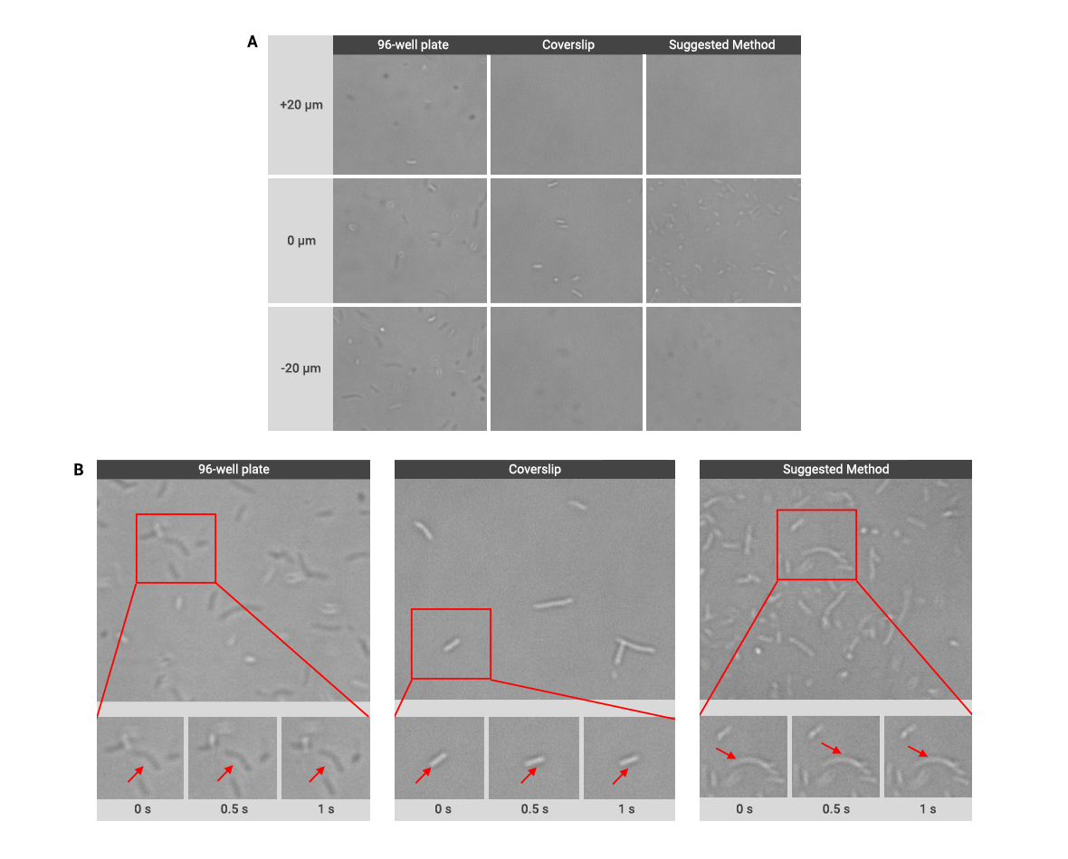
Figure 1. Montages captured from different z-planes of -20 μm, 0 μm, and +20 μm (A) and Brownian motion within 1 second (B) using traditional methods with a 96-well plate and a coverslip on a glass slide, and the suggested method.
An assessment was conducted to evaluate the image quality obtained through conventional methods using a 96-well plate and a coverslip on a glass slide, in comparison to our suggested method. Significant advantages were observed when using our method compared to traditional methods. Utilizing bright-field (BF), fluorescence (FL) images, and their overlay images for assessment, our method exhibited superior performance (Figure 2). Notably, focus issues and misalignment between BF and FL images were notably reduced in the overlay images obtained with the suggested method. This enhancement in image quality highlights our method’s effectiveness in minimizing artifacts and promoting more precise alignment, thus offering researchers improved visual clarity and more reliable imaging results for their investigations.
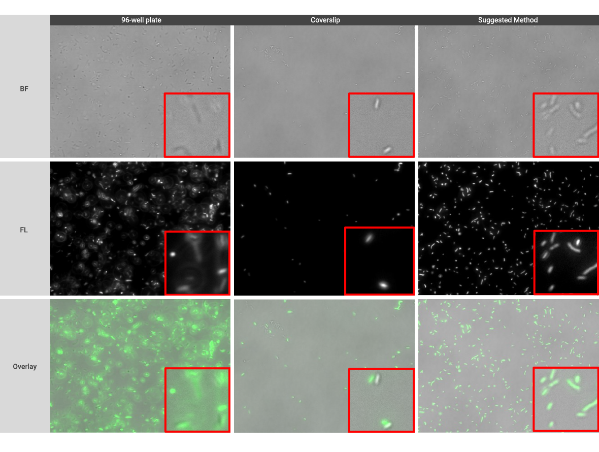
Figure 2. A montage shows the comparison of imaging quality between traditional methods with a 96-well plate and a coverslip on a glass slide, and the suggested method.
We have demonstrated the significant improvement in bacteria image quality achieved by combining the sample preparation method for use with the QUANTOM Tx™ Microbial Cell Counterand the CELENA® X High Content Imaging System for high-resolution imaging. This innovative approach addresses the challenges posed by Brownian motion and z-plane scattering which yields remarkable improvements in image quality compared to traditional methods. Consequently, this strategy offers a straightforward and efficient approach to achieve superior bacterial imaging.


