3D Cell Culture: Differences Between Spheroids and Organoids and Their Analytical Strategies Core Platforms for 3D Cell-Based Disease Modeling and Drug Screening
Spheroids and organoids are three-dimensional (3D) cell culture models developed to overcome the limitations of conventional two-dimensional (2D) cell culture systems. These models have become essential platforms in life science and biopharmaceutical research, enabling tumor microenvironment modeling, stem cell–based tissue reconstruction, and drug response evaluation.
Spheroids represent relatively simple three-dimensional cellular aggregates, whereas organoids are more advanced models in which stem cells or tissue-derived cells undergo self-organization to partially recapitulate the structural and functional characteristics of native organs.
In this article, we provide a scientific overview of the definitions, formation principles, applications, and analytical strategies of spheroids and organoids, with particular emphasis on the importance of 3D imaging and quantitative cell analysis technologies.
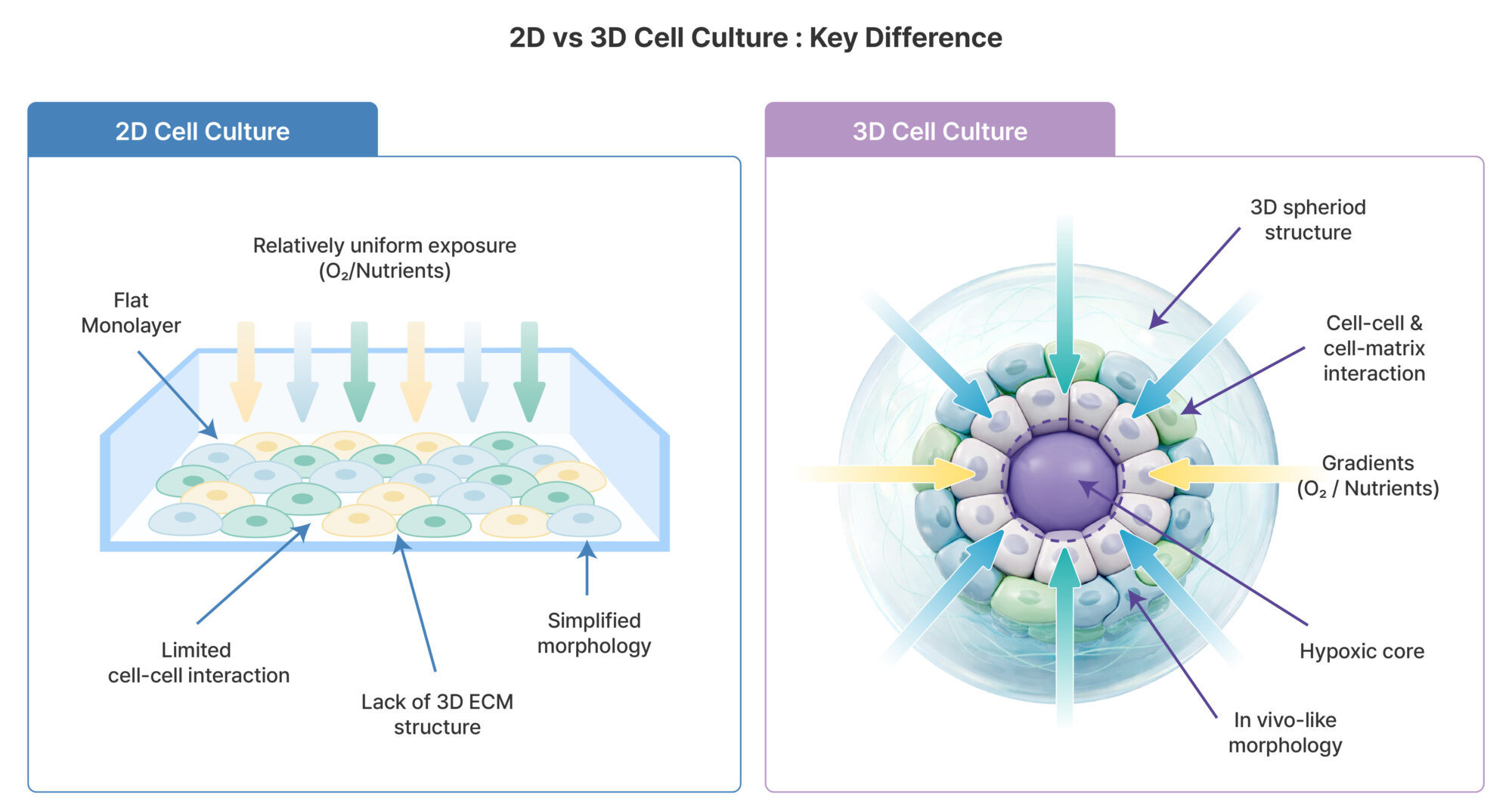
Figure 1. Structural differences between 2D and 3D cell culture systems In 2D culture systems, cells grow as a flat monolayer and are exposed to relatively uniform levels of oxygen and nutrients. In contrast, 3D culture systems enable cell–cell and cell–extracellular matrix (ECM) interactions, leading to the formation of oxygen and nutrient gradients and the development of hypoxic regions in the core. These structural differences play a critical role in more accurately recapitulating the in vivo microenvironment.
1. Why 3D Cell Culture?
Conventional two-dimensional (2D) monolayer culture systems have inherent limitations in fully recapitulating cell–cell and cell–matrix interactions. In 2D environments, cells grow attached to an artificial flat surface, making it difficult to reproduce the spatial organization, diffusion limitations, and microenvironment-driven signaling observed in native tissues.
In cancer research, anticancer drug screening, and stem cell–based disease modeling, the ability to mimic the in vivo microenvironment is directly linked to the accurate interpretation of experimental outcomes.
Three-dimensional (3D) culture systems offer several key advantages:
Maintenance of cell polarity
Formation of oxygen and nutrient gradients
Representation of differential drug penetration
Gene expression patterns more closely resembling those of native tissues
Owing to these characteristics, spheroids and organoids have evolved beyond simple cellular aggregates and are now recognized as essential platforms for enhancing physiological relevance in preclinical research.
2. Spheroids: Simple Yet Powerful 3D Tumor Models
▷ Definition
A spheroid is a three-dimensional, spherical cellular aggregate formed by the spontaneous assembly of one or more cell types without attachment to a substrate. In particular, multicellular tumor spheroids (MCTS) are widely used as representative 3D models that recapitulate key aspects of the tumor microenvironment and are extensively applied in anticancer drug evaluation and tumor biology research.
▷ Formation Methods
Ultra-low attachment plates
Hanging drop method
Spinner flasks
Microfluidic systems
These approaches minimize cell adhesion to artificial surfaces and promote spontaneous cell aggregation, enabling the formation of uniformly sized spheroids.
▷ Biological Characteristics
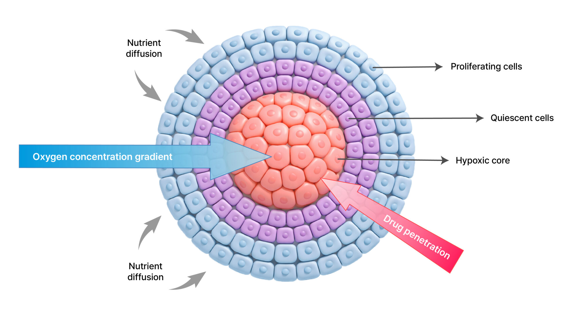
Within a spheroid, distinct structural regions are typically established:
Outer layer: proliferating cells
Intermediate layer: quiescent cells
Central region: hypoxic/necrotic core
In this three-dimensional structure, the diffusion of oxygen and nutrients is restricted, leading to the formation of concentration gradients toward the core. As a result, hypoxic conditions develop in the central region, and in larger spheroids, necrosis may occur.
Similarly, therapeutic agents diffuse from the outer layers toward the center, often exhibiting reduced penetration into the core. This diffusion limitation serves as an experimental model for therapeutic resistance mechanisms observed in solid tumors.
Consequently, spheroid-based analyses provide a more physiologically relevant platform than 2D culture systems for evaluating drug efficacy during the preclinical stage.
▷ Major Applications
Evaluation of anticancer drug efficacy
Assessment of radiosensitivity
Studies of tumor invasion and metastasis
Immune response analysis through co-culture with immune cells
Figure 2. Structural characteristics and gradient formation in tumor spheroids Tumor spheroids are typically composed of an outer proliferating layer, an intermediate quiescent zone, and a central hypoxic and/or necrotic core. Due to limitations in oxygen and nutrient diffusion, concentration gradients are established toward the center of the spheroid. As a result, therapeutic agents may not sufficiently penetrate into the core region. These structural characteristics are critical for understanding differential drug responses observed between 2D and 3D tumor models.
3. Organoids: Stem Cell–Derived Models that Recapitulate Organ Structure
▷ Definition
An organoid is a three-dimensional cell model in which pluripotent stem cells (PSCs) or adult stem cells undergo self-organization within a 3D environment to form structures that partially recapitulate the architecture and function of native organs.
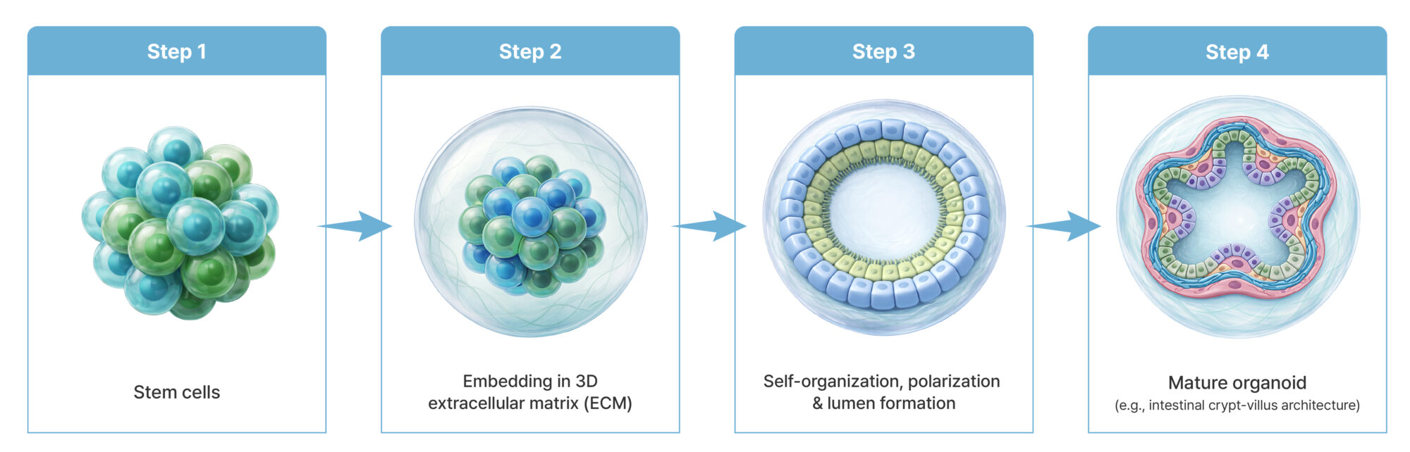
Figure 3. Formation of organoids from stem cells When cultured within a three-dimensional extracellular matrix (ECM) environment, stem cells undergo self-organization, polarization, and lumen formation, ultimately forming organoids with organ-specific architecture. During this process, cell–cell interactions and ECM-mediated signaling act in concert, leading to progressive increases in structural complexity. The resulting organoids partially recapitulate both structural and functional characteristics of native tissues.
▷ Representative Examples
Intestinal organoids
Brain organoids
Liver organoids
Tumor organoids (patient-derived organoids, PDOs)
▷ Key Characteristics
Category
Spheroid
Organoid
Cellular Composition
Single or mixed cell populations
Stem cell–derived multicellular structures
Structural Complexity
Relatively simple
Organ-specific architecture
Functional Recapitulation
Limited
Partial reproduction of organ-specific functions
Applications
Tumor models
Disease modeling, regenerative medicine
Organoids extend beyond simple three-dimensional cell aggregates by reflecting organ-specific microarchitecture and cellular differentiation patterns. Notably, certain organoid systems can partially replicate functional features such as barrier function, secretory activity, metabolic processes, and electrophysiological properties. This enables their application in functional disease modeling, going beyond purely morphological analysis.
Furthermore, patient-derived organoids (PDOs) can preserve individual tumor or tissue characteristics, making them valuable platforms for precision oncology and personalized therapeutic strategy development.
4. Key Considerations in 3D Cell Model Analysis: Imaging & Quantification
To accurately evaluate three-dimensional (3D) structures, quantitative image-based analysis is essential, going beyond conventional viability assays.
▷ Major Analytical Parameters
Spheroid diameter and volume
Morphological changes
Live/dead cell ratio
Invasion distance
Fluorescence intensity gradients
High-resolution fluorescence imaging combined with automated cell analysis systems plays a critical role in reducing experimental variability and ensuring reproducible data acquisition.
Due to the structural complexity of 3D models, imaging depth and optical clarity are particularly important. To address these challenges, tissue-clearing techniques or confocal microscopy–based imaging approaches are commonly employed.
Furthermore, automated data extraction using quantitative analysis software is essential in high-throughput screening environments.
▷ High-Content Imaging Approaches for 3D Analysis
To meet the demands of quantitative 3D analysis, automated imaging systems capable of multi-focal plane (Z-stack) acquisition are highly effective. For example, the CELENA® X High Content Imaging System enables automated Z-stack acquisition, capturing depth-resolved images of spheroids and organoids that can be integrated for quantitative analysis.
This approach minimizes structural information loss associated with single focal-plane imaging and supports consistent measurement of morphological parameters such as spheroid area, circularity, and growth kinetics. When combined with fluorescence-based live/dead assays, it allows quantification that reflects the entire 3D structure, thereby improving the accuracy of organoid viability assessment.
In particular, automated imaging and analysis within multi-well plate formats reduce inter-experimental variability and provide reproducible datasets suitable for drug screening and comparative drug response studies.
Therefore, in 3D cell model research, imaging and quantitative analysis should be regarded not as auxiliary procedures but as integral components of experimental design.
5. Spheroid vs. Organoid: Selecting the Appropriate Model Based on Research Objectives
▷ For Drug Screening
→ Spheroid models are suitable for applications requiring rapid and highly reproducible evaluation.
▷ For Disease Modeling and Functional Analysis
→ Organoids are more appropriate when structural complexity and organ-specific characteristics are required.
▷ For Precision Medicine
→ Patient-derived organoids (PDOs) enable drug response assessment tailored to individual patients. Selecting the appropriate 3D model requires careful consideration of research objectives, cost, experimental duration, and available analytical infrastructure.
▶ Example 1: Anticancer Drug Testing
In some cases, lower IC₅₀ values have been observed in 2D culture systems, whereas reduced drug efficacy has been reported in spheroid models due to limited drug penetration. This highlights the ability of 3D models to more realistically reflect therapeutic responses observed in vivo.
▶ Example 2: Intestinal Organoids
Intestinal organoids form crypt–villus–like structures and recapitulate barrier function as well as stem cell niche organization. These characteristics make them valuable models for studying inflammatory bowel disease (IBD) and host–microbiome interactions.
6. Future Perspectives of 3D Cell Culture
Integration with organoid-on-a-chip technologies
Development of multi-organoid systems
Advanced AI-based image analysis
Automation of high-content screening
Personalized drug response prediction
3D cell culture technologies are evolving beyond simple structural modeling, integrating with microfluidics-based organoid-on-a-chip platforms and multi-organoid systems to achieve greater physiological sophistication. These advanced platforms provide a foundation for more accurately reflecting inter-organ communication and drug dynamics.
At the same time, quantitative data generated through high-resolution imaging are increasingly combined with AI-driven analytical algorithms, enabling large-scale data processing and advanced pattern recognition. In the future, image-derived morphological parameters from 3D models are expected to be integrated with genomic and transcriptomic (omics) data, further improving the accuracy of disease-specific response prediction and personalized drug response assessment.
Such technological advancements are anticipated to position 3D cell-based assays not merely as complementary tools to traditional 2D systems, but as standard preclinical platforms that simultaneously ensure physiological relevance and predictive accuracy.
Conclusion
Spheroids and organoids represent core platforms within 3D cell culture technology, and their importance continues to grow across various fields, including cancer research, disease modeling, drug development, and personalized therapeutic strategies.
In particular, the integration of high-precision imaging with automated analytical systems significantly enhances data reliability and research efficiency in 3D models.
Looking ahead, 3D cell-based assays are expected to become standard platforms in preclinical research rather than auxiliary models.
Logos Biosystems provides cell analysis and imaging solutions for a wide range of life science research applications.
Friedrich J, Seidel C, Ebner R, Kunz-Schughart LA. Spheroid-based drug screen: considerations and practical approach. Nat Protoc. 2009.
Edmondson R, Broglie JJ, Adcock AF, Yang L. Three-dimensional cell culture systems and their applications in drug discovery and cell-based biosensors. Assay Drug Dev Technol. 2014.
Lancaster MA et al. Cerebral organoids model human brain development and microcephaly. Nature. 2013.
Clevers H. Modeling development and disease with organoids. Cell. 2016.