The History of Automated Cell Counting: A Journey from Coulter Counters to Image-Based Cell Counters
Automated cell counting technology has significantly transformed the field of life sciences, revolutionizing the way researchers count and analyze cells. While we explore the future trends and innovations shaping this field, it is important to acknowledge the historical milestones that laid the foundation for automated cell counting. In this blog, we will not only delve into the latest advancements but also take a journey through the history of automated cell counters, including the invention of the Coulter counter, the introduction of the flow cytometry, and the impact of image-based cell counters. By understanding the historical context, we gain a deeper appreciation for the progress made and the possibilities that lie ahead.
The Invention of the Coulter Counter
The history of automated cell counting can be traced back to the invention of the Coulter counter by Wallace H. Coulter in the 1950s. The Coulter counter introduced the principle of electrical impedance to cell counting, enabling rapid and accurate measurements. This innovative technology relies on the detection of changes in electrical resistance as cells pass through a small aperture. By analyzing the changes in impedance, the Coulter counter can determine the number and size distribution of cells in a sample. This groundbreaking invention revolutionized cell counting, making it faster, more precise, and less labor-intensive.
The Coulter counter, despite its significant contributions to automated cell counting, has certain limitations that are important to consider. Firstly, it struggles to count clustered cells effectively due to its fundamental principle of counting objects passing through an orifice as individual cells. As a result, the Coulter counter has primarily been utilized for counting non-clustering blood cells. In typical laboratory settings, where a majority of cells are attached cells, the process of cell suspension often leads to the formation of cell clusters. Consequently, the Coulter counter faces challenges in accurately counting such cells, limiting its applicability in routine cell culture laboratories.
Additionally, the Coulter counter cannot measure cell viability. The assessment of cell viability often requires the use of dyes, such as trypan blue, which can distinguish between cells that have taken up the dye and those that haven’t, allowing for viability to be determined. Unfortunately, the Coulter counter lacks the capability to differentiate viability based on such dye uptake.
These limitations have led to the Coulter counter being predominantly employed for diagnostic purposes in counting blood cells, while its utilization in research laboratories has been limited. Researchers often require techniques that can effectively count clustered cells and provide insights into cell viability, prompting the exploration of alternative automated cell counting technologies to address these specific research needs.
The Introduction of the Flow Cytometry
Flow cytometry is a powerful technique that has revolutionized cell analysis and cell counting. Developed in the 1960s, flow cytometry allows for the rapid analysis of individual cells by suspending them in a fluid stream and passing them through a laser beam. As cells pass through the beam, they scatter light and emit fluorescence, providing information about their size, granularity, and molecular characteristics. Flow cytometers can measure multiple parameters simultaneously, enabling researchers to analyze complex cell populations. The integration of automated sample handling systems with flow cytometry has further enhanced its efficiency and throughput, making it an indispensable tool in fields such as immunology, cancer research, and drug discovery.
Flow cytometry has been widely used as a sophisticated instrument for cell analysis including cell counting and viability measurement. However, it has had several important drawbacks:
Firstly, flow cytometry primarily measures fluorescence, making it incapable of measuring cell viability using commonly used methods like trypan blue staining in the laboratory. Secondly, due to its complexity, flow cytometry required specialized operators, making quick and simple cell counting challenging. Most significantly, flow cytometry was an expensive instrument, typically used in core facilities rather than individual laboratories, limiting its accessibility.
These limitations have hindered the widespread adoption of flow cytometry for routine cell counting and viability assessment in many laboratory settings.
The Impact of Image-Based Cell Counters
The revolutionary impact in the history of automated cell counting is the development of image-based cell counters based on automated microscopy. In the early 2000s, advancements in digital imaging technology and computer algorithms allowed researchers to automate the cell counting process using microscopic images. These systems employ sophisticated image analysis algorithms to identify and count cells based on their morphological features, such as size, shape, and color. Image-based cell counters provide enhanced accuracy and the ability to analyze various cell types, making them invaluable tools in cell culture workflows and research applications. Here are critical aspects of the image based cell counters that impact cell counting process.
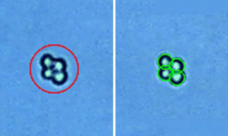
Distinguishing Clustered Cells:
One of the major advantages of image-based cell counters is their ability to accurately count clustered cells. Unlike other methods that treat clustered cells as a single entity, image-based counters leverage advanced imaging techniques to distinguish individual cells within clusters. This capability improves counting accuracy, especially in samples where cell clustering is prevalent, such as suspension cultures or tissue samples.
Advanced Cluster Analysis
Versatile Staining Methods:
Image-based cell counters can utilize a variety of staining methods. Stains like
trypan blue and fluorescence dyes such as
acridine orange/propidium iodide (AOPI) can be employed to measure viability. This versatility in staining methods allows researchers to choose optimized methods based on their cell types and sample conditions.
Advanced Image Analysis Algorithms:
With sophisticated image analysis algorithms, image-based cell counters can identify and exclude non-cellular objects from the counting process. This includes debris, dirt particles, or artifacts that may be present in the sample or counting chamber. By eliminating these non-cellular elements, the counters ensure more accurate and reliable cell counting results.
Compact and Affordable Instrumentation:
Modern image-based
cell counters feature compact optical systems, making them more affordable and accessible to a wider range of users. These instruments are designed to be compact and benchtop-sized, requiring minimal laboratory space. The affordability and compactness of these devices have made them a viable option for various research settings, including small laboratories or field studies.
Conclusion
The field of automated cell counting has undergone significant transformation over the years. The journey from the invention of the Coulter counter to the advent of flow cytometry and image-based cell counters reveals the impressive strides made in technology and its impact on life sciences. Each evolution has addressed limitations of its predecessors, offering improved precision, versatility, and reliability.
The introduction of image-based
cell counters however, marked a revolutionary leap forward. By utilizing advanced imaging technology and computer algorithms, these counters overcome many limitations of previous technologies, offering the ability to accurately count clustered cells, use versatile staining methods, and exclude non-cellular objects for more reliable results. Moreover, their compact and affordable design has democratized access to this vital technology, making it more available to various research settings.
As we appreciate the historical milestones and current capabilities of automated cell counting, we also eagerly anticipate future advancements in this field. The marriage of cell counting technology with AI, machine learning, and potentially, novel biosensors, promises a future of even greater precision and functionality. The evolution of automated cell counting is far from over; it continues to shape and empower life sciences research, making way for exciting discoveries and advancements in the years to come.
Overall, cell viability measurement is a crucial technique in toxicology studies. By accurately measuring the viability of cells, researchers can identify the potential toxicity of substances, evaluate their effects on living organisms, and develop effective treatment strategies to minimize their impact.
Logos Biosystems provides a range of image-based cell counters, catering to different needs, from affordable benchtop cell counters to advanced bacterial cell counters. For more information, please visit
www.logosbio.com.