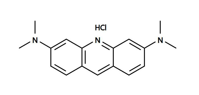
Acridine Orange (AO): AO is a small molecule with a molecular weight of 265 Da that can penetrate both live and dead cells. Once inside a cell, it selectively binds to nucleic acids and emits a distinctive green fluorescence. When AO is bound to DNA, it has a peak excitation at approximately 500 nm and peak emission at 525 nm.
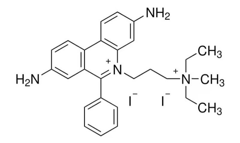
Propidium Iodide (PI): PI has a molecular weight of 668 Da and cannot penetrate the live cell membrane. PI can penetrate membrane compromised cells such as dying or dead cells. When PI is bound to DNA, it has peak excitation at 535 nm and peak emission at 617 nm, and as a result, emits a distinctive red fluorescence.
When AO/PI mixture is used to stain cells, live cells emits green fluorescence and dead cells emits red fluorescence when cells are excited with proper light source. In detail, AO can penetrate all cells and binds do nucleic acids, while PI can only penetrate dead cells and then binds to nucleic acids in dead cells. Since live cells allow AO penetration only, the fluorescence of PI is not observed in live cells. In dead cells, both dyes can penetrate and theoretically both AO and PI fluorescence can be observed. However, due to the FRET phenomenon, where AO emission is absorbed to PI, only the fluorescence of PI is selectively observed in dead cells. This means that the fluorescence of AO is not observed in dead cells when AO and PI are used together. Using a fluorescence microscope or an automatic cell counter that adopts the principle of a fluorescence microscope, green fluorescence of AO is observed in living cells, while red fluorescence of PI is observed in dead cells.

1. Preparing Your Cells:
① Start by harvesting your cells from the culture.
② Collect the cells using the centrifuge, then resuspend them in PBS.
2. The Staining Process:
① Prepare 10x AO/PI staining solution (E.g., Acridine Orange/Propidium Iodide Stain, Cat # F20331, Logos Biosystems).
② Mix 18 uL cell suspension with 2 uL , Acridine Orange/Propidium Iodide Stain. AO/PI stains cells immediately, and no further incubation is necessary.
3. Cell Counting
Using fluorescence microscope:
① Load 10 uL of stained cells into the hemocytometer.
② Using a fluorescence microscope fitted with the appropriate filters, bring your cells into focus. The colors will be evident: count green fluorescent cells for the live cells and and red fluorescent cells for the dead cells.
③ To determine cell concentration, count all cells in five large squares of the hemocytometer grid.
Cell concentration per mL=(average number of cells per square/volume of one square (0.1 μL) x dilution factor
✓ If 10x AO/PI is used, the dilution factor is 1.11
④ To determine cell viability, use this formula:
Percentage viability=(Number of green fluorescent cells/Total number of cells)×100
Using the automated fluorescence cell counter (LUNA-FLTM),(LUNA-FX7TM)
① Load 10 uL of stained cells into the cell counting chamber slide (E.g., PhotonSlide, Cat# L12005)
② Insert the chamber slide onto the cell counter and press “Count”
③ Automated cell counter will display all necessary information automatically such as cell concentration, call viability, and cell size, etc.
When measuring cell viability using Acridine Orange/Propidium Iodide (AO/PI) staining, several key points need to be kept in mind to ensure accurate results. Here are some essential tips to remember:
Consistency in Dye Concentration: The concentration of AO and PI should be consistent across experiments to obtain comparable results. Any changes in dye concentration could impact staining intensity and the differentiation between live and dead cells.
Protection from Light: Both AO and PI are photosensitive. To prevent the degradation or photobleaching of the dyes, always handle and store them in the dark or under dim light conditions.
Timing: The staining process with AO/PI is relatively quick. Prolonged exposure of cells to the dyes can lead to inaccurate results. It’s essential to keep the incubation time consistent between samples.
Sample Handling: Gentle mixing of the cell suspension with the dyes is essential to prevent cell damage and to ensure homogeneous staining. However, avoid vigorous shaking or pipetting that can harm the cells.
Freshness of Dye Solutions: Check the shelf-life of dye solutions. Over time, the dyes can degrade or become contaminated, which could affect their staining efficacy.
Calibration and Control:When you are using the automated cell counter, ensure your cell counter is calibrated with the control samples. For control samples, use known concentration of fluorescent bead mixture (E.g., LUNA-FX Calibration Beads Kit, Cat# F73101) or Cell Counter Validation Slide-FL (Cat# L72030)
Other Methods for Confirmation: It’s always a good idea to validate AO/PI staining results with another method, especially if you observe unexpected results or if the results have significant implications.
Remember, while AO/PI staining is a useful and rapid technique for assessing cell viability, it’s essential to maintain a standardized procedure to obtain accurate and reproducible results
For top-quality reagents assured for cell viability measurement, visit www.logosbio.com.