Since the late 1990s, a series of image-based automated cell counters have been introduced to provide accurate cell number and viability data. Basically, these cell counters use light microscopy and computer-based image analysis software. Instead of the human eye used to count manually, magnified cell images are automatically captured by a digital camera and transported to the computer where various software algorithms are used to accurately find cells and discriminate against non-cellular objects such as cell debris.
To determine cell viability, dye-exclusion methods have been widely used. Popular dyes used for cell viability are trypan blue and methylene blue. Because stained dead cells are easily discriminated using bright field imaging, this technique is successfully used in most automated cell counters including the LUNA™ automated cell counter(see “How to Count Cells: An Overview of Cell Counting Methods” for general review) (https://logosbio.com/application_notes/how-to-count-cells-an-overview-of-cell-counting-methods).
Although bright field image analysis has been successfully applied to most mammalian cell counting, a lot of cell or sample types remain challenging. For example, primary cells and peripheral blood mononuclear cells (PBMCs) are recognized as difficult-to-count cells with the bright field image analysis. Typically, these cells are mixed with a large population of red blood cells, which can be mistakenly counted as dead cells in bright field image analysis. Other challenging cell samples include: yeast cells due to their small size and sperm cell because of regular impurities.
To overcome the current limitations of bright field image based cell counting, more sensitive and accurate cell counting methods are necessary. Fluorescence cell counters such as the LUNA-FL™ automated cell counter, the LUNA-FX7™ automated cell counter is a recently introduced fluorescence cell counter used to count cells without the limitations of cell types.
Fluorescence (FL) is a term to describe the emission of light by a substance that has absorbed light. The substance becomes excited and unstable when light is absorbed and tries to go back to its ground state by emitting energy in the form of light. In most cases, the emitted light has a longer wavelength, i.e., lower energy, than the absorbed radiation (Fig 1 and Fig 2).
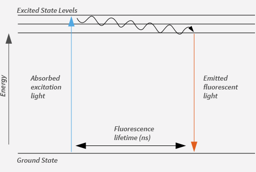
Figure 1. A diagram showing the principle of fluorescence from Pierce homepage.
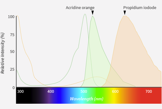
Figure 2. Spectra of Acridine Orange and Propidium Iodide from Invitrogen homepage.
FL dyes for nucleic acid staining can be classified based on their physical,chemical and fluorescent characteristics as summarized in Table 1.
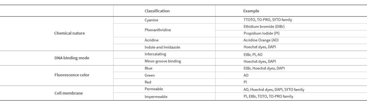
Table 1. Classification of nucleic acid-binding FL dyes
Important factors to be considered for fluorescence cell counting include 1) cell membrane permeability, 2) fluorescence spectrum and 3) fluorescence strength. Because cell membranes consist of lipid bilayers containing long stretches of hydrocarbon fatty acid tails, they act as hydrophobic barriers of cells and have selective permeability to various molecules. In general, small, non-polar FL dyes can readily cross the cell membrane and stain nucleic acids whereas bulky, polar, especially charged dyes cannot. Acridine Orange (AO) and Propidium Iodide (PI) are good examples of cell membrane permeable and impermeable FL dyes, respectively (Fig 3).
Cell membrane permeable FL dyes (e.g., AO) can stain nucleic acids of both live and dead cells so that they are able to visualize the entire population. Cell membrane impermeable FL dyes (e.g., PI) can only enter and visualize only dead cells in which membrane integrity is compromised. Therefore, combining AO/PI dual staining (https://logosbio.com/cell_counting_acc/acridine-orange-propidium-iodide-stain/) and automated cell counting system enables the efficient distinction between live and dead cells as well as accurate determination of their numbers.
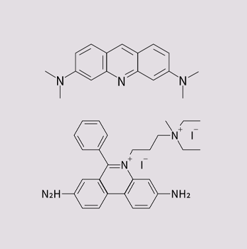
Figure 3. Chemical structure of Acridine orange(upper) and Propidium iodide(lower)
Fluorescence-based automated cell counting is useful in various applications where the bright field-based counterpart is not feasible. Counting White Blood Cells (WBCs) in peripheral blood is a good example (Fig 4). Blood cells are significantly small in size (~6mm) for bright field-based cell counting. To make it worse, WBCs are intermingled with the vast majority of anucleated Red Blood Cells (RBCs) and platelets. Automated cell counting using AO/PI dual staining automatically excludes RBCs and platelets from the total cell count. Because mature RBCs and platelets lose their nuclei during the differentiation, they do not stain with either dye. Manually tedious, time-consuming and error-prone WBC counting can be performed effortlessly with the fluorescence-based automated cell counter. Counting PBMCs in the presence of contaminating RBCs and platelets is another example (Fig 5).
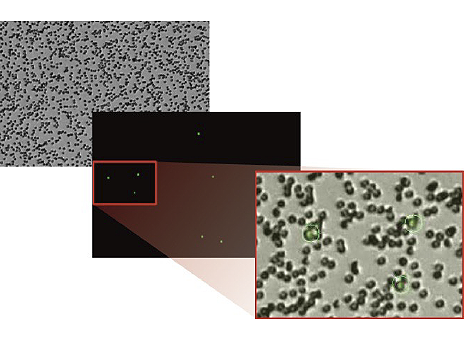
Figure 4. Counting human peripheral blood with Luna-FL and the AO/PI kit.
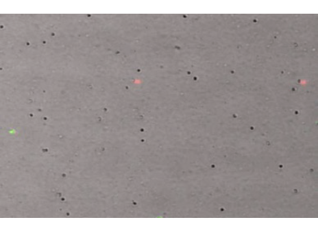
Figure 5. Counting mouse peripheral blood mononuclear cells (PBMCs) with Luna-FL and the AO/PI kit.
To date, two main technologies have been adopted to count fluorescently labeled cells: flow cytometry and image-based analysis. Flow cytometry is one of the most powerful tools to analyze cells using a fluorescence detection system. Regular flow cytometers are not adequate for determining cell numbers because they do not measure the volume of liquid passed while fluorescently labeled cells are being counted. Only volumetric flow cytometry can count the absolute number of cells in unit volumes of liquid to provide highly accurate cell numbers that multiple researchers have proven comparable to those obtained with hemocytometers (1 – 4). Conventional flow cytometers can be used to estimate the absolute cell number if FL beads with a known number are mixed with experimental samples with an unknown number (5). The cell numbers obtained from the bead-based estimation and the direct volumetric flow cytometry are not statistically different (6).
Despite the advantages of flow cytometric cell counting, the instruments are relatively expensive with costs ranging from $40,000 to over $100,000. Because of the complicated optical, fluidic and electro-mechanical configuration, users need to take special training before using it or core facilities need to hire a professional operator. Moreover, continuous maintenance is required for the acquisition of reliable data, which includes de-clogging, optical alignment and laser adjustment. For these reasons, they are not widely used for general cell counting applications.
The concept of image-based cell counting was implemented by combining fluorescence microscopy and computer software for digital image analysis (7, 8 and 9). To better fit the need for automatic cell counting, dedicated cell counters have started to come into the market, which are equipped with a fluorescence microscope module and an image analyzing/counting algorithm. The current trend is that the instruments are designed in a small, all-in-one configuration so that they can be employed in space-limited environments, and the instrument/consumable price is affordable by most laboratories.
When compared to flow cytometers, image-based cell counters(https://logosbio.com/compare-cell-counters/) are not as versatile as their flow-based counterparts, but they have more capability than required for general purpose cell counting. They are more affordable and easier to learn than the flow-based system, and essentially maintenance-free. Another noteworthy advantage over flow cytometry is that cells being counted can be visually inspected so users can verify counting accuracy in real-time. The presence of any cellular debris and abnormal cell morphology can be checked as well.
Currently, a number of fluorescence cell counters are commercially available ranging from simple (a single fluorescence channel) to comprehensive models (up to seven fluorescence channels). They differ in their hardware (size, measuring volume, built-in monitor and counting principle; image- or flow-based), software (speed, de-clustering and image cytometry capability), list price and running cost (the price of consumables per count).
Fluorescence cell counters such as the LUNA-FL™ automated cell counter(https://logosbio.com/luna-fl/) , the LUNA-FX7™ automated cell counter(https://logosbio.com/luna-fx7/) have been developed for sensitive and accurate live/dead cell counting results without limitation of sample types. For cell counting and viability analysis, AO/PI dye mixture is provided as a reagent kit (AO/PI cell viability kit, Cat# F23001)(https://logosbio.com/cell_counting_acc/acridine-orange-propidium-iodide-stain/). After the sample is stained with AO/PI cell viability kit, it is loaded onto the disposable cell counting chamber named PhotonSlide™. The PhotonSlide™ is made of ultra-low auto-flurescence plastic material, which leads to more sensitive cell detection. The LUNA fluorescence cell counters captures and analyzes multi color images, bright field, green fluorescence, and red fluorescence. Within 30 seconds, total cell numbers, viability, cell size and histogram data are displayed on a touch screen LCD monitor. The LUNA-FL™ is the most affordable fluorescence cell counter on the market in terms of both the price of instrument and disposables. In addition, the Luna FX7 automated cell counter is optimized for bioprocess operations in the production of cell therapy or gene therapy products, and is currently the most precise and accurate automated cell counter developed to date. It also includes software that complies with the essential specifications of 21CFR part 11, which is required for equipment used in GMP facility. In summary, the LUNA series is a cell counter based on sensitive and accurate fluorescence staining methods to provide cost-effective and reliable cell counting solutions.


