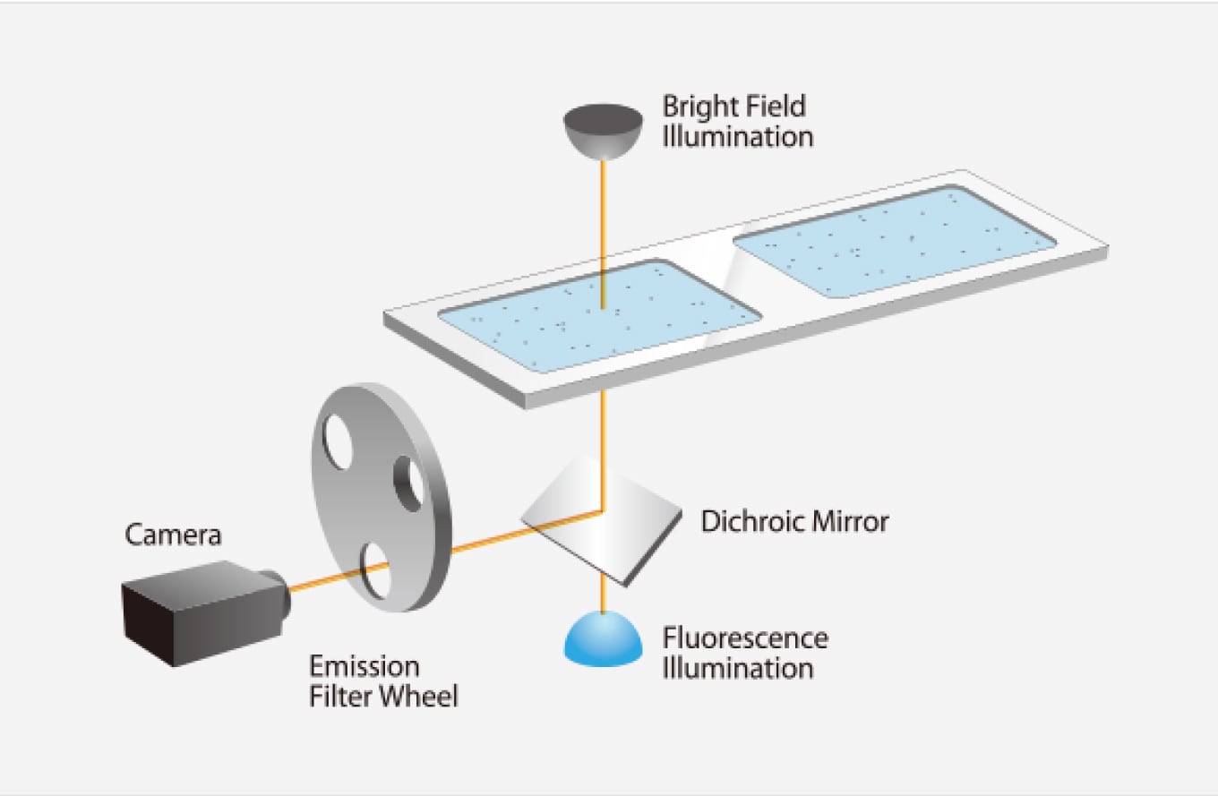
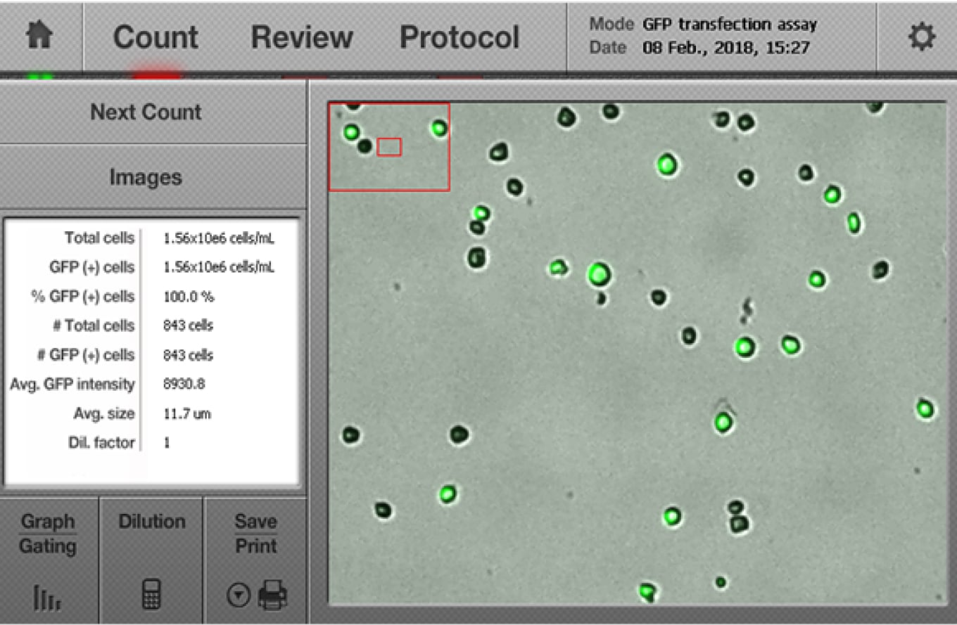
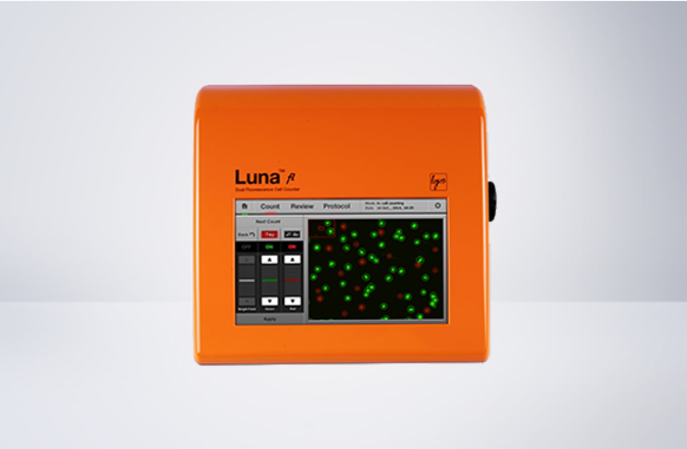
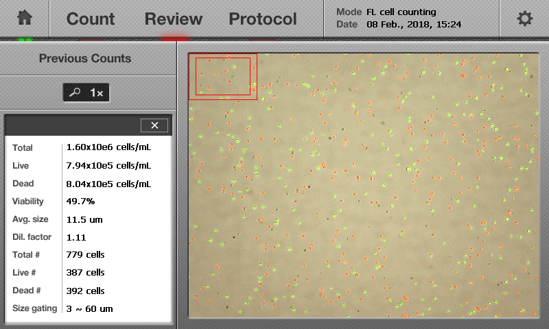
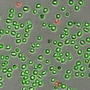
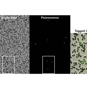
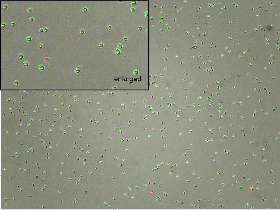
LUNA-FL™は、デュアル蛍光および明視野光学系を搭載した高性能セルカウンターです。細胞の種類や大きさに制限されることなく、細胞数、細胞生存率、そしてGFP形質転換効率を評価できます。
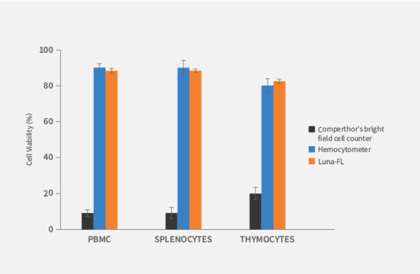
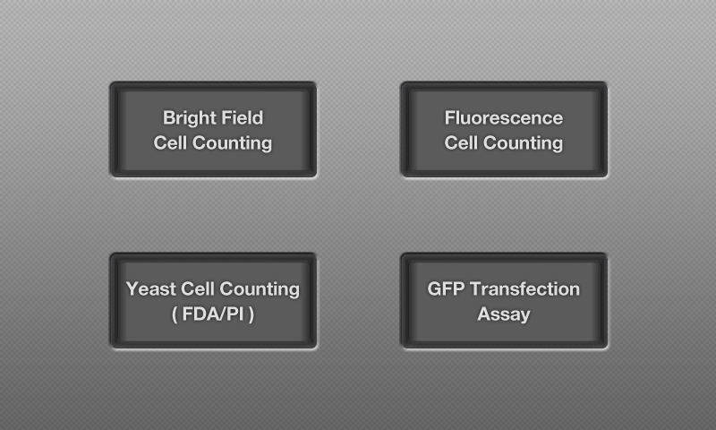
4つのカウントモードを備えたLUNA-FL™は、まさに当社のフラッグシップモデルです。明視野およびデュアル蛍光光学系を搭載し、細菌を除くほとんどの細胞タイプを高感度で検出可能です。PBMC、脾臓細胞、好中球、幹細胞などの初代細胞を不要なデブリと区別し、正確な細胞数と生存率を測定します。明視野セルカウント、蛍光セルカウント、酵母セルカウント、GFP形質転換アッセイなど、多彩な用途に対応。コンパクトなデザインで、細胞培養フード内やベンチトップでも快適に使用でき、ワークフローにスムーズに組み込めます。

Great performance, would recommend for other researchers!
Easy to use, saves us time and offers good quality. The consultant Dr Eberhardt was also professional and patient to teach us before use.
University Heidelberg
Great instrument, I like using it. Really helpful.
The device is easy to use, I mainly use it before cell seeding or to check for my culture's viability and survival. The results are reproducible and the device provides fast results. The accuracy seems to be great as well.
BGU
Brain Cancer Cell-derived Exosomes Protect Scopolamine-Induced Death of SH-SY5Y Neuron Cells.
2021. Lee M. International Journal of Health Sciences and Research 10.36838/v3i1.8.
The Type and Source of Reactive Oxygen Species Influences the Outcome of Oxidative Stress in Cultured Cells.
2021. Goffart S, Tikkanen P, Michell C, Wilson T, Pohjoismäki JLOLO. Cells 10(5):1075.
Immune Memory in Mild COVID-19 Patients and Unexposed Donors Reveals Persistent T Cell Responses After SARS-CoV-2 Infection.
2021. Ansari A, Arya R, Sachan S, Jha SN, Kalia A, Lall A, Sette A, Grifoni A, Weiskopf D, Coshic P, Sharma A, Gupta N. Frontiers in Immunology 12:636768.
The Small GTPase Arf6 Functions as a Membrane Tether in a Chemically-Defined Reconstitution System.
2021. Fujibayashi K, Mima J. Frontiers in Cell and Developmental Biology 9:628910.
Flow cytometric quantification of apoptotic and proliferating cells applying an improved method for dissociation of spheroids.
2021. Metzger W, Rösch B, Sossong D, Bubel M, Pohlemann T. Cell Biology International 10.1002/cbin.11618.
2022-04-07 | 2.12 MB
2022-04-07 | 2.14 MB
2018-04-16 | 110 KB
2018-04-16 | 284 KB
2018-04-16 | 1.7 MB
2018-04-13 | 906 KB
2018-04-16 | 403 MB
2018-04-16 | 862 KB
2018-04-16 | 476 KB
2018-04-13 | 665.84 KB
2018-04-13 | 720.79 KB
| Cat # | Product | Qty |
|---|---|---|
| L20001 | LUNA-FL™ Automated Fluorescence Cell Counter | 1 unit |
| L12005 | PhotonSlide™, 50 Slides | 1 box |
| L12006 | PhotonSlide™, 500 Slides | 10 boxes |
| L12007 | PhotonSlide™, 1000 Slides | 20 boxes |
| L12001 | LUNA™ Cell Counting Slides, 50 Slides | 1 box |
| L12002 | LUNA™ Cell Counting Slides, 500 Slides | 10 boxes |
| L12003 | LUNA™ Cell Counting Slides, 1000 Slides | 20 boxes |
| L12011 | LUNA™ Reusable Slide | 1 unit |
| L12012 | LUNA™ Reusable Slides | 2 units |
| L12014 | LUNA™ Reusable Slide Coverslips | 10 units |
| T13001 | Trypan Blue Stain, 0.4% | 2 X 1 mL |
| L13002 | Erythrosin B Stain | 2 x 1 mL |
| F23001 | Acridine Orange/Propidium Iodide Stain | 2 x 0.5 mL |
| F23002 | Acridine Orange Stain | 2 x 0.5 mL |
| F23003 | Propidium Iodide Stain | 2 x 0.5 mL |
| F23202 | Yeast Viability Kit 1 – Propidium Iodide Stain for Yeast (1 x 0.5 mL) – Fluorescein Diacetate Stain (1 x 0.5 mL) – Cell Dilution Buffer (1 x 20 mL) – Fluorescence Signal Enhancer 1 (1 x 0.5 mL) |
1 kit |
| F23212 | Cell Dilution Buffer | 5 x 20 mL |
| F23102 | LUNA™ Fluorescence Calibration Beads | 1 x 0.5 mL |
| B13101 | LUNA™ Standard Beads | 2 x 1 mL |
| F24003 | LUNA-FL™ IQ/OQ Protocol | 1 copy |
| P10001 | LUNA™ Printer I | 1 unit |
| P12001 | LUNA™ Printer I Paper (Thermal, 275 prints/roll, 6rolls) | 3 x 2 rolls |
| Sample Volume | 10 μl |
| Cell Counting Time | 30 sec |
| Cell Concentration Range | 5×10⁴ – 1×10⁷ cells/mL |
| Cell Size Range | Detectable Range: 1 – 90 μm Optimal Range: 5 – 60 μm |
| Excitation wavelength | 470 ± 20 nm |
| Emission wavelength | 530 ± 25 nm, 600 nm (LP) |
| Light Source | LED |
| Image Resolution | 5 MP |
| LCD Display | 800 x 480 pixels |
| Dimensions (W×D×H) | 22 × 21 × 9 cm (8.6 × 8.3 × 3.5 in) |
| Weight | 1.8 kg (4 lb) *without the AC adapter and power cord |


