Many common procedures and assays require that scientists first obtain an accurate count of the number or density of cells. Such procedures range from simple cell culture maintenance, such as cell splitting, to quantitative experiments such as qPCR. Counting cells can be done in a number of ways, and in this article we seek to provide a comprehensive overview of how to count cells using the various cell counting methods, as well as the benefits and drawbacks of each method.
To use a hemocytometer, it should first be cleaned with 70% ethanol and lens paper. The coverslip is then gently placed atop the counting chamber. You will know the coverslip is properly positioned if you can observe a phenomena known as Newton’s rings; a concentric-ring pattern of color. A small sample of cell suspension is taken using a pipette and the pipette is placed near the edge of the chamber, allowing the cell suspension to enter the counting chamber by capillary action. If cell viability is to determined, trypan blue should be added to the cell suspension (in a 1:1 ratio) prior to addition to the chamber.
Counting chambers, more frequently referred to as hemocytometers, were the first method developed specifically for obtaining accurate cell counts. A special, gridded chamber is affixed to a glass slide. Generally, the squares measure 1 mm x 1 mm and are further subdivided into 0.05 mm x 0.05 mm squares. The chamber’s edges are designed to hold a special glass coverslip 0.1 mm above the marked grid, thereby creating demarcated areas of known volume.
The microscope is then focused on an area of the counting chamber and the cells are counted using a tally counter. This is typically done using the 1 mm2, 100 nl area of the counting chamber and a 4x or 10x objective, but the precise area and objective used will depend on the size of your cells and their density in suspension. This process is generally repeated using four different 1 mm2 areas and the results are averaged. If determining cell viability, separate counts should be made for live and dead cells, with the dead cells appearing blue due to the permeability of their damaged membranes to trypan blue.
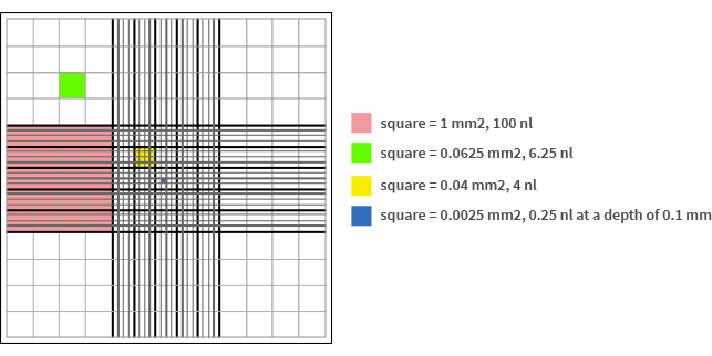
Fig 1 Schematic representation of a hemocytometer grid.
Benefits & Drawbacks: Manual cell counting is the least expensive method of determining cell count and cell density, however it is also the slowest and most tedious. It can also be one of the least reliable due to the possibility for human error, particularly if performing many cell counts sequentially, and performing a large number of cell counts can also cause eye strain. If a lab’s needs to count cells is sporadic or infrequent, and if a high degree of precision in the cell count is not required, then hemocytometers can be a good choice. For more frequent use, when greater accuracy is required, or in higher-throughput applications, counting chambers fall short. Additionally, human counters are often poor at discerning between multiple types of cells in a suspension unless the differences in size and / or shape between the various cell types are extreme.
Automated cell counters were designed to be a faster, easier, automated alternative to manual counting. They use the same principles of operation as hemocytometers; they perform multiple counts of cells within a known area and average out the results. They also can discern live cells from dead cells using dye exclusion methods (such as trypan blue). Automated cell counters may either operate as standalone devices or require a connection to a computer. In addition to a cell count, most counters also provide statistical information on cell size. Benefits & Drawbacks: Manual cell counting is the least expensive method of determining cell count and cell density, however it is also the slowest and most tedious. It can also be one of the least reliable due to the possibility for human error, particularly if performing many cell counts sequentially, and performing a large number of cell counts can also cause eye strain. If a lab’s needs to count cells is sporadic or infrequent, and if a high degree of precision in the cell count is not required, then hemocytometers can be a good choice. For more frequent use, when greater accuracy is required, or in higher-throughput applications, counting chambers fall short. Additionally, human counters are often poor at discerning between multiple types of cells in a suspension unless the differences in size and / or shape between the various cell types are extreme.
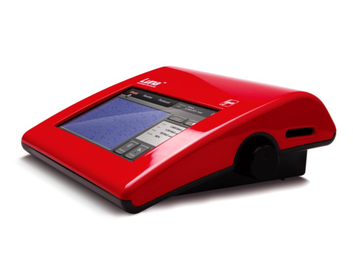
Fluorescent cell counters, such as the Luna-FL™ dual fluorescence cell counter, distinguish live and dead cells as well as perform a cell count by using common stains such as acridine orange and propidium iodide. These types of cell counters are also superior at counting cells in cultures which may be contaminated with non-cell debris, such as primary cells, as the dyes will clearly distinguish between cells. Benefits & Drawbacks: For general purpose cell counting and cell viability applications, automated cell counters are an affordable and high-throughput solution. The cost of operation is low, they are easy to use, and they greatly reduce the amount of human effort required to count cells. They are both precise and reliable, but may have difficulty obtaining accurate measurements of cells that are highly irregularly shaped, are extremely small, or are in cell suspensions that are extremely dilute or contain a large variety of cells that need to be distinguished. For most cell types and most applications, automated cell counters provide excellent counting performance at a relatively low cost.
Coulter counters are not optical instruments, but rather measure the electrical resistance across one or more microchannels. Cells, having greater resistance than the electrolyte solution that the cells are suspended in, cause a brief increase in resistance when passed between the channels. This change, which increases with cell size, is detected by the Coulter counter. Use of a Coulter counter is somewhat similar to that of an automated cell counter. The cell suspension is diluted, as needed, thoroughly mixed to ensure an even cell distribution, added to a vial, and a run is started. Unlike automated cell counters, however, use of Counter counters requires running a blank first and also flushing the device after use. Benefits & Drawbacks: Because of their relative speed compared to manual counting and their ability to accurately count cells of differing size, Coulter counters are frequently used for complete blood counts in hospitals, where red blood cells and white blood cells need to be quickly and accurately distinguished. However, Coulter counters are not capable of distinguishing live cells from dead cells, nor do they accurately count cells which form clusters or clumps. They also require more maintenance.
Flow Cytometers are most frequently used for more detailed cellular analysis, as they are equipped with fluorescence detection technologies that can detect labelled intracellular components. Not all flow cytometers are capable of determining cell count, as not all draw specific liquid volumes or measure the volume of liquid drawn. Those that do, however, are capable of providing highly accurate cell counts, and can discriminate cells based on factors such as protein expression by using fluorescently labelled antibodies. This makes them able to discern cell types of the same size within the same sample, or even the same types of cells at different cell stages. However, such complex experiments also complicate the experimental set up and antibody incubations are often on the scale of hours, thereby potentially extending the workflow quite significantly. Use of a flow cytometer is quite simple (load and run), and therefore the overall ease of use depends more heavily on the experimental set-up. Benefits & Drawbacks: Flow cytometers are extremely powerful cellular analysis tools but are also extremely expensive, with costs ranging from $40,000 to over $100,000. Because of this, they are rarely used for general cell counting applications.
Spectrophotometry is occasionally used to obtain relative estimates of cell density. Since cells are turbid, less light will pass through the cuvette as the cell density increases. However, since spectrophotometers do not actually count cells but rather measure absorbance, and since other variable components of cell suspensions can effect absorbance, spectrophotometers are not a reliable method of estimating cell density. To estimate cell density using a spectrophotometer, place the cell suspension in a cuvette and measure the absorbance. If you are looking to get a relative density measurement, you may simply compare to another sample. Otherwise, you must compare to cell suspensions of known density in order to estimate an absolute cell density. Plating is another method of counting cells, although only in colony-forming cells such as bacteria. To count cells with plating, the cells are heavily diluted and streaked onto a plate. After given sufficient time for colony growth, the number of colonies are counted. Based on the dilution and the known volume of suspension that was streaked onto the plate, the density of the original suspension can be determined. Plating is only a useful method for microbes, and due to the time required for colony formation it is also the slowest method.


