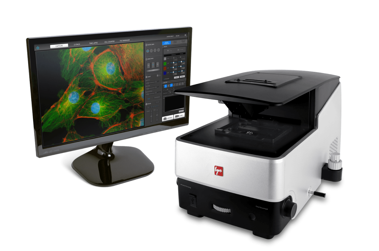
Desde la obtención de imágenes de fluorescencia multicolor hasta el análisis de datos en un único equipo
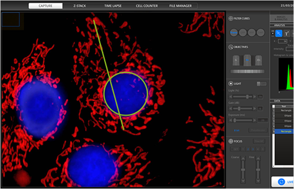
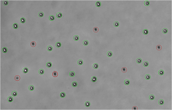
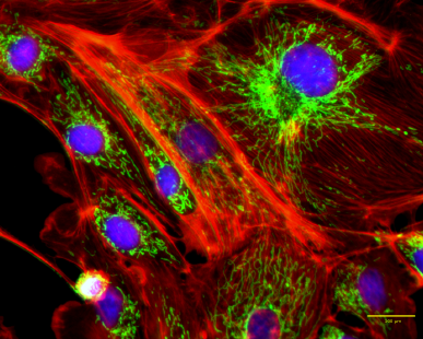
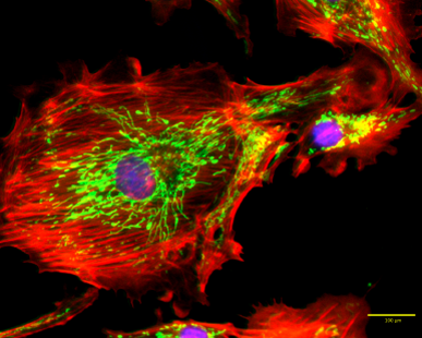
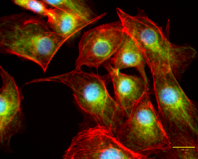
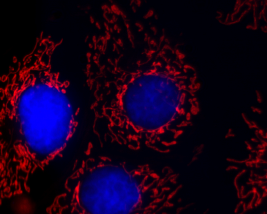
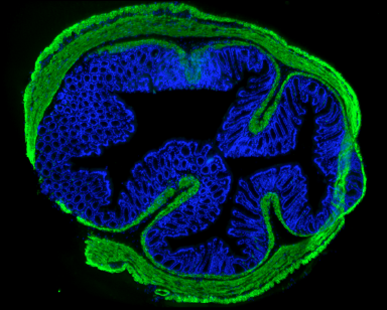
Con el sistema de captura de imágenes digitales CELENA® S puede capturar fácilmente imágenes de alta resolución, con calidad de publicación. El CELENA® S le sorprenderá con su pequeño tamaño ya que cuenta con una óptica de precisión avanzada, un sensor CMOS, filtros LED fluorescentes digitalmente controlados, todo ello con un procesador y un software de análisis integrado. El sofisticado pero sencillo software permite obtener imágenes de fluorescencia multicolor, imágenes de campo claro, imágenes de contraste de fase, imágenes de células vivas en un lapso de tiempo e imágenes apiladas en Z (Z-stack).
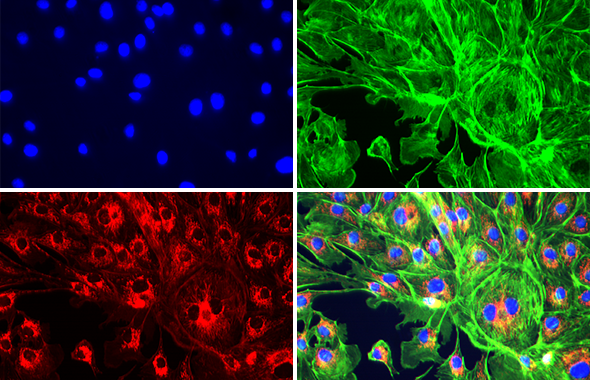

Con el sistema de incubación CELENA® S on-stage, puede realizar una gran variedad de experimentos para la obtención de imágenes de células vivas en condiciones fisiológicas y no fisiológicas. El mezclador de gases permite un control preciso de la composición del aire, la temperatura y la humedad. Un controlador de temperatura calienta la tapa de la incubadora y la placa por separado para que pueda regular la temperatura del sistema y evitar la condensación.
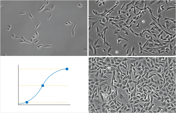
CELENA® S permite configurar fácilmente las secuencias de imágenes en un lapso de tiempo, así como exportar las imágenes y crear vídeos que incluyan anotaciones.


It is an essencial for every cell culture lab.
The equipment is very is to use to all its potential, with minimal training. It allows high resolution images of publishing quality.
Dental Medicine, U. Porto
Great equipment. This microscope was excellent value and is simple to use. If you use a vibration isolation table, it is perfectly designed for eliminating the complexities of microscopy without compromise of performance and the Celena makes cell imaging gives optimum results. The Celena imaging system is just right for cell culture.
GPR171 Activation Modulates Nociceptor Functions, Alleviating Pathologic Pain.
2021. Cho PS, Lee HK, Choi YI, Choi SI, Lim JY, Kim M, Kim H, Jung SJ, Hwang SW. Biomedicines 9(3):256.
Rheological properties of cellulose nanofiber hydrogel for high-fidelity 3D printing.
2021. Shin S, Hyun J. Carbohydrate Polymers, 263:117976.
Novel regulatory roles of UCP1 in osteoblasts.
2021. Mukherjee S, Yun JW. Life Sciences 276:119427.
Human WRN is an intrinsic inhibitor of progerin, abnormal splicing product of lamin A.
2021. Kang SM, Yoon MH, Lee SJ, Ahn J, Yi SA, Nam KH, Park S, Woo TG, Cho JH, Lee J, Ha NC, Park BJ. Scientific Reports 11(1):9122.
Inhibition of Lipopolysaccharide-Induced Inflammatory and Oxidative Responses by Trans-cinnamaldehyde in C2C12 Myoblasts.
2021. Park C, Lee H, Hong S, Molagoda IMN, Jeong JW, Jin CY, Kim GY, Choi SH, Hong SH, Choi YH. International Journal of Medical Sciences 18(12):2480-2492.
2026-04-24 | 939 KB
2026-04-24 | 3 MB
2021-06-18 | 389.25 KB
2018-04-17 | 526 KB
2018-04-17 | 644 KB
2018-04-17 | 958 KB

| Cat # | Product | Qty |
|---|---|---|
| CS20001 | CELENA® S Digital Imaging System | 1 unit |
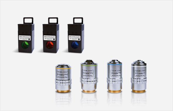
| CS20002 | CELENA® S Digital Imaging System Starter Kit – 4 Objectives – 3 LED Filter Cubes |
1 unit |
| I10520 | CS Stage Top Incubator [Tokai] | 1 set |
| I10501 | Universal Heating System [Ibidi] | 1 set |
| I10502 | Gas Incubation System for CO2 [Ibidi] | 1 set |
| I10503 | Gas Incubation System for CO2/O2 [Ibidi] | 1 set |
| I10201 | Universal Holder | 1 unit |
| I10202 | 25 mm x 75 mm Slide Holder, Two Positions | 1 unit |
| I10203 | 35 mm Cell Culture Dish Holder, Four Positions | 1 unit |
| I10204 | 60 mm Cell Culture Dish Holder, Two Positions | 1 unit |
| I10205 | 100 mm Cell Culture Dish Holder, One Position | 1 unit |
| I10206 | 25 c㎡ Nunc T-25 Flask Holder, Two Positions | 1 unit |
| I10207 | 75 c㎡ Nunc T-75 Flask Holder, One Position | 1 unit |
| I10208 | 25 c㎡ BD/Greiner T-25 Flask Holder, Two Positions | 1 unit |
| I10209 | 75 c㎡ BD/Greiner T-75 Flask Holder, One Position | 1 unit |
| I10210 | Glass Hemocytometer Holder, One Position | 1 unit |
| Imaging methods | Epifluorescence and transmitted light (brightfield and phase contrast) |
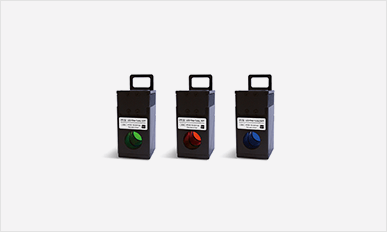
| Illumination | LED filter cubes with adjustable intensity (>50,000 hr life per filter cube) |
| Fluorescence channels | 3 fluorescence channels and 1 transmitted light channel |
| Objective turret | 5 positions |
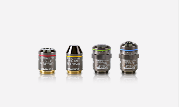
| Objectives | High quality long working distance (LWD) and coverslip-corrected; 1.25X-100X |
| Condenser | 47 mm LWD condenser; 3-positions with brightfield and phase contrast annuli |
| Computer | Built-in dual core CPU, 128 GB SSD internal storage |
| Stage | Mechanical X/Y stage, motorized Z stage; accommodates an onstage incubator |
| LCD display | Full HD color LCD monitor, 1920 x 1080 pixels (not included) |
| Camera | 1.3 MP monochrome CMOS with 1280 x 1024 pixels |
| Images | 8 or 16-bit TIFF, JPG, BMP, or PNG |
| Dimensions (L x W x H) | 44 cm x 30 cm x 27 cm (17.3 x 11.6 x 10.6 in) |
| Weight | 20 kg (44 lb) |


