Keywords
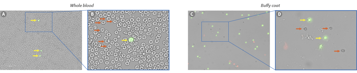
Peripheral Blood Mononuclear Cells (PBMCs) are a vital source material used in myriad research applications, including single-cell sequencing to vaccine development and toxicological studies. Furthermore, PBMC derivatives such as T cells, B cells, NK cells, and stem cells are based on cell therapies, including CAR-T cell therapies and regenerative medicines. Therefore, accurately measuring numbers and viability of PBMCs after collection, isolation, or expansion are essential to making experimental or manufacturing decisions about downstream processes. Yet, directly obtaining leukocyte counts in whole blood using traditional counting techniques is complicated by the presence of mature, anucleated RBCs and platelets. Advantageously, the use of nucleic acid stains like Acridine Orange and Propidium Iodide (AO/PI) allow the nucleated leukocytes to be differentiated and accurately counted within a whole blood sample (Figure 1). Here, we exhibit the ability of the new dual fluorescent LUNA-FX7™ Automated Cell Counter to meet diverse cell counting needs by demonstrating its use in counting leukocytes in whole blood and the PBMCs isolated from whole blood samples.
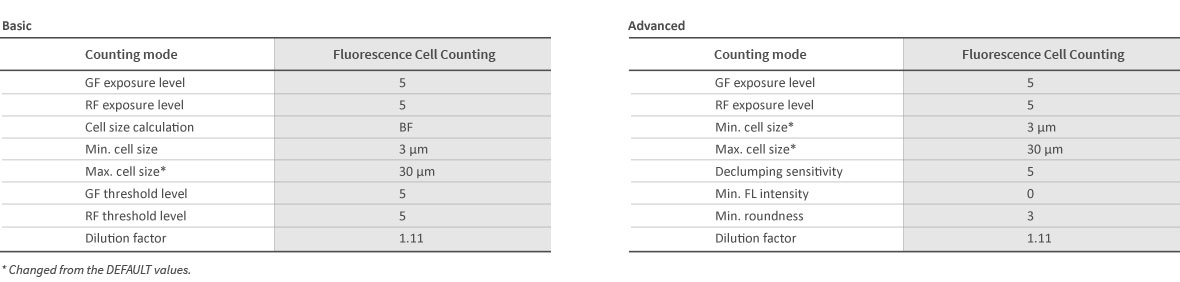
One milliliter of the human peripheral blood sample was prepared, and the PBMC sample was obtained by standard density gradient centrifugation technique using Histopaque-1083 (Sigma, #10831)1. After final washing, the PBMCs enriched in the buffy coat were resuspended in 100 µl PBS or RPMI +10% FBS media. Cell counts were performed in the LUNA-FX7™ with either the 2-channel PhotonSlide™ (Cat# L12005) or LUNA™ 8-Channel Slides (Cat# F72001) and used a modified default protocol in the Fluorescence Cell Counting mode (Table 1). Before loading the cells, cells were stained at the standard ratio of the AO/PI reagent (Cat# F23001); 18 µl of cells + 2 µl of AO/PI, and then 10 µl of the mix was loaded into a slide chamber.
Evaluating the PBMC concentration and viability with the LUNA-FX7™
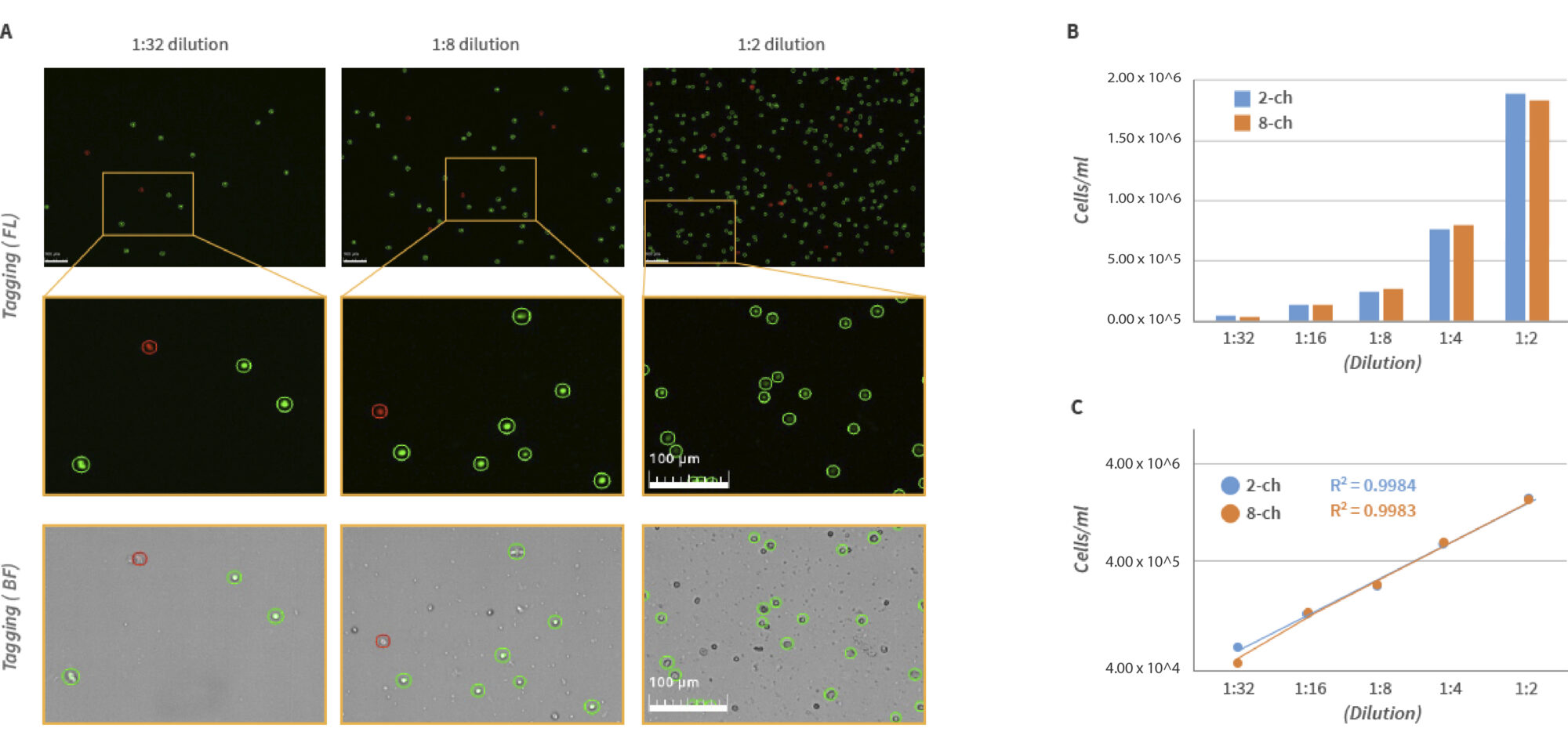
The separated PBMCs were analyzed by counting a series of 2-fold dilutions using both the 8-channel and 2-channel slides (Figure 2). The counts using both slide types showed linearity with an R-square value of 0.99 or above on the logarithmic scale of concentrations over the serial dilutions. Not surprisingly, with a greater volume of analysis, the 2-channel slide showed slightly better consistency than the 8-channel slide.
Table 1. The optimized parameter settings for PBMC or leukocytes counting of the LUNA-FX7™ on Fluorescence Cell Counting mode
The leukocytes in whole blood were visualized by fluorescence and also accurately enumerated with the LUNA-FX7™. Among countless mature RBCs, the fluorescence leukocytes were distinctively counted from diluted whole blood (Figure 3).
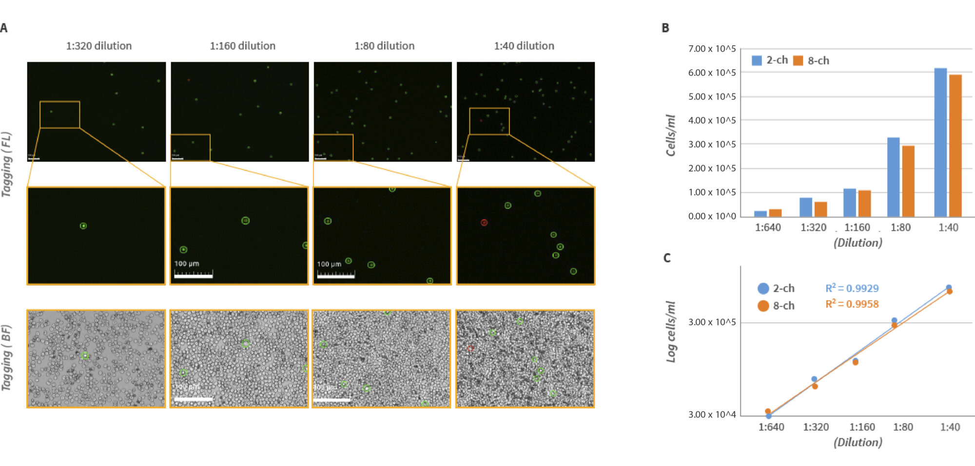
The enumeration of PBMCs was evaluated adequately on the LUNA-FX7™ Automatic Cell Counter with the 2-channel PhotonSlide™ and LUNA™ 8-Channel Slides. With more slide options for the LUNA-FX7™, LUNA™ 1-Channel Slides (Cat# L72011) may be applied for a more comprehensive concentration range by analyzing 47 image fields, and LUNA™ 3-Channel Slides (Cat# L72021) are beneficial for preset of triplicate analysis as counting options. Indeed, leukocyte counting is much more straightforward by loading the diluted blood after AO/PI fluorescence staining. So, you can use the LUNA-FX7™ to respect the archived records of PBMCs along with leukocyte counts without intensive labor.