Keywords
Bacteria quantification is a one of the most fundamental measurements in a number of fields, including healthcare, agriculture, food production, and industry. From the need to accurately quantify live Lactobacillus casei in probiotics, heat-killed Vibrio cholera in a vaccine, sulfur oxidizing bacteria for waste treatment, general bacterial levels to determine soil contamination, or just your standard cell suspension in a laboratory, bacterial cell counting is a basic but significant step. Here, we compare three common bacterial detection and quantification methods with an rapid and automated solution to bacterial quantification.
One of the most common methods to quantify bacteria is counting colony forming units (CFUs). This widely used method is simple, gives a good general idea of cell viability, and is sensitive even to low concentrations of bacteria. However, this method is dependent on growth media and conditions as well as being limited to culturable bacteria, which excludes viable but not culturable (VBNC) strains. Another major disadvantage is that it takes days to get results that are estimations at best. One colony may arise from one or a thousand cells and sample preparation can vary from tech to tech, as well as each time, depending on sample conditions. The time investment is a significant drawback when assessing time-sensitive issues such as food or water contamination concerns.
Specialty hemocytometers (Petroff-Hausser and Levy counting chambers) are used to directly count bacteria under a microscope. This gold standard of cell counting is a relatively rapid and easy way to get results, but it is labor intensive and prone to user-to-user variability. The minute size of bacteria mean that, not only are some species difficult to see even under a microscope, they can move within the counting chamber as well as being distributed across different focal planes, making it difficult to assess and control whether counts are done accurately in a reproducible way each time.
Flow cytometry is recognized as one of the most accurate ways to determine total and viable cell counts. On the other hand, flow cytometry requires extensive training to operate the instrument as well as analyze the data. Similar to colony counting, a significant limitation of flow cytometry is that it cannot distinguish between a single cell or a cluster of cells. Each particle is registered as a single event, so cocci in clusters or bacilli in chains cannot be identified as single cells.
The QUANTOM Tx™ Microbial Cell Counter is an image-based, automated cell counter that can identify and count individual bacterial cells in minutes. The QUANTOM Tx™ automatically focuses in on, captures, and analyzes multiple images of fluorescence-stained cells to detect bacterial cells with high sensitivity and accuracy. The sophisticated cell detection and declustering algorithm that can accurately identify individual bacterial cells in even the tightest clusters. Utilizing two different stains, the QUANTOM Tx™ can count total cells or viable cells. Stained cells are mixed with QUANTOM Tx™ Cell Loading Buffer I, loaded into QUANTOM Tx™ M50 Cell Counting Slides, and spun in the QUANTOM Tx™ Centrifuge to immobilize and evenly distribute the cells along a single focal plane to ensure accurate cell detection. Counting results and images can be viewed and saved immediately after the count.
Counting with the QUANTOM Tx™ Microbial Cell Counter
The counting accuracy of the QUANTOM Tx™ was confirmed by counting standard fluorescent beads of known concentration. Beads were serially diluted, mixed with the QUANTOM™ Cell Loading Buffer I, and loaded into a QUANTOM™ M50 Cell Counting Slide. The slide was spun in the QUANTOM™ Centrifuge at 300 RCF for 10 minutes to ensure even bead distribution and then counted with the following parameters with the QUANTOM Tx™ : LED = Bead, size = 1-50 μm, detection sensitivity = 0, declustering level = 7, roundness = 30 %. All counts were performed in triplicate. As demonstrated in Figure 1, the QUANTOM Tx™ produced results with a a strong correlation (R2 = 0.9997) to the theoretical concentration of beads.
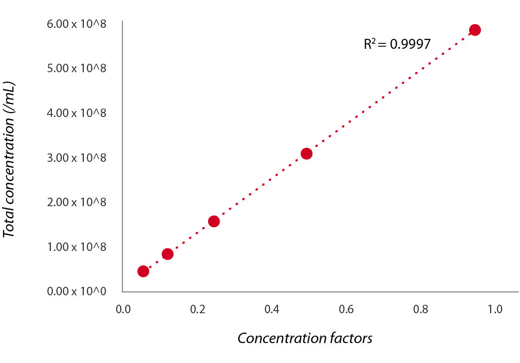
Figure 1. Correlation between known standard bead concentrations and QUANTOM Tx™ results.
To compare the accuracy of the QUANTOM Tx™ compared to a flow cytometer and hemocytometer, serial dilutions of Escherichia coli were counted in triplicate. For the flow cytometer, cells were stained with Thiazol Orange and counted with the FACSCalibur flow cytometer (BD Biosciences). Standard beads were added to help determine cell concentration. For the hemocytometer, cells were loaded into a Petroff-Hausser Counting Chamber (20 μm, Hausser Scientific) and imaged with the CELENA® S Digital Imaging System (Logos Biosystems) using a TC PlanAchro 20x Ph objective. Five squares of the Neubauer counting grids were counted. For the QUANTOM Tx™, cells were stained with QUANTOM™ Total Staining Dye and counted with the following parameters: LED = 5, size = 0.3-50 μm, detection sensitivity = 0, declustering level = 0, roundness = 0 %. There was no significant difference in the total concentrations, but the hemocytometer showed a higher variability from count to count as cell concentration increased whereas the QUANTOM Tx™ and flow cytometer had more consistent results (Figure 2).
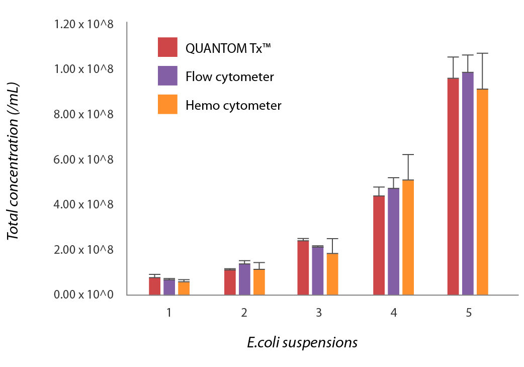
Figure 2. Comparison of counting results from the QUANTOM Tx™, a flow cytometer, and a hemocytometer.
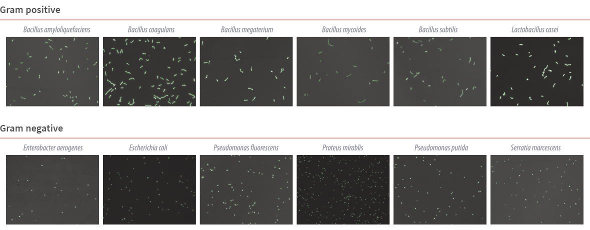
To demonstrate the versatility of the QUANTOM Tx™, various bacteria species were stained with QUANTOM™ Total Staining Dye and counted with the QUANTOM Tx™. The QUANTOM Tx™ automatically captures, analyzes, and labels up to 20 high-resolution images per count, making it easy to verify the accuracy of each count visually. Lactobacillus casei were counted with the following parameters : LED = 5, size = 1-50 μm, detection sensitivity = 0, declustering level = 7, roundness = 0. Bacillus megaterium were counted with the following parameters: LED = 5, size = 1-50 μm, detection sensitivity = 3, declustering level = 10, roundness = 0. A more complete list of bacteria and the parameters used to count them can be found at: goo.gl/ixr8kr. As shown in Figure 3, the QUANTOM Tx™ was able to distinguish individual cells in the tight bacilli chains.
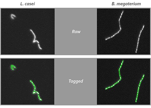
Figure 3. Individual bacilli in chains detected using the QUANTOM Tx™ declustering feature. Lactobacillus casei (L) and Bacillus megaterium (R) were counted with the QUANTOM Tx with the declustering levels set to 7 and 10, respectively.
Bacteria are an incredibly diverse group of organisms that come in a variety of shapes, sizes, and arrangements, making automated cell counting a challenging feat. The ubiquitous colony counting method is a time-consuming, unreliable estimation at best. Even flow cytometers register each particle, single or clustered, as a single event. Hemocytometers are time consuming and prone to user-to-user variability. The QUANTOM Tx™ Microbial Cell Counter is an effective, accurate, and rapid way to quantify bacteria of various morphologies and arrangements. This makes the QUANTOM Tx™ an important and necessary tool for immediate, real-time assessment of the bacterial population within a sample.
A partial list of bacteria tested on the QUANTOM Tx™


