Keywords: SpectraSlide® AP-1, LUNA-FX7™ automated cell counter, cell viability, pipette-free, stain-integrated
Automated cell counters, like the LUNA-FX7™, have revolutionized cell counting by replacing traditional labor-intensive methods. However, the process still requires additional labware and staining dyes, which can be challenging for beginners and those with limited access to laboratory resources. To address these limitations, we developed the SpectraSlide® AP-1, a user-friendly disposable cell counting slide designed to simplify workflows while ensuring accuracy. Given its unique approach to cell counting, validating its accuracy and reproducibility is essential. In this study, we conducted a comparative analysis of SpectraSlide® AP-1 and 포톤슬라이드 (PhotonSlide™), as 포톤슬라이드 (PhotonSlide™) is already widely used as a standard for cell counting. Using U937 and PBMC samples, we assessed their linearity in measuring cell concentration and viability to validate the precision and effectiveness of the SpectraSlide® AP-1.
Introducing the SpectraSlide® AP-1
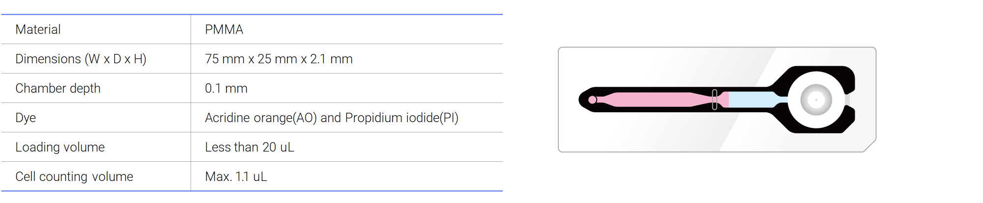
The SpectraSlide® AP-1 is an innovative disposable cell counting device designed for the LUNA-FX7™ automated cell counter. Traditional cell counting methods often require multiple tools, such as pipettes and external staining reagents, adding complexity to the workflow. The SpectraSlide® AP-1 simplifies this process by integrating cell sampling and staining into a single, efficient step.
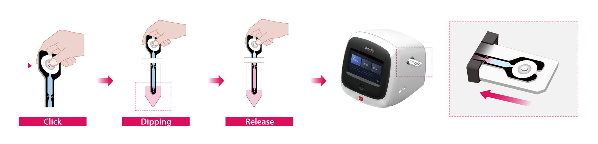
Its pre-coated acridine orange (AO) and propidium iodide (PI) stain solution provides an extended shelf life at room temperature. Upon contact with liquid, the stain dissolves instantly, allowing for immediate and uniform cell staining without any additional mixing. These dyes efficiently penetrate and label nuclei, providing accurate and reproducible viability assessments. Additionally, the slide’s user-friendly sampling button facilitates effortless sample loading, making cell counting more consistent and accessible for researchers. By reducing handling steps and ensuring precise viability measurements, the SpectraSlide® AP-1 enhances workflow efficiency while maintaining high accuracy and reliability.
Human peripheral blood was processed to obtain a PBMC sample using the standard density gradient centrifugation technique with Histopaque-1083 (Sigma, #10831). After the final wash, PBMCs enriched in the buffy coat were resuspended in PBS containing 1% BSA.
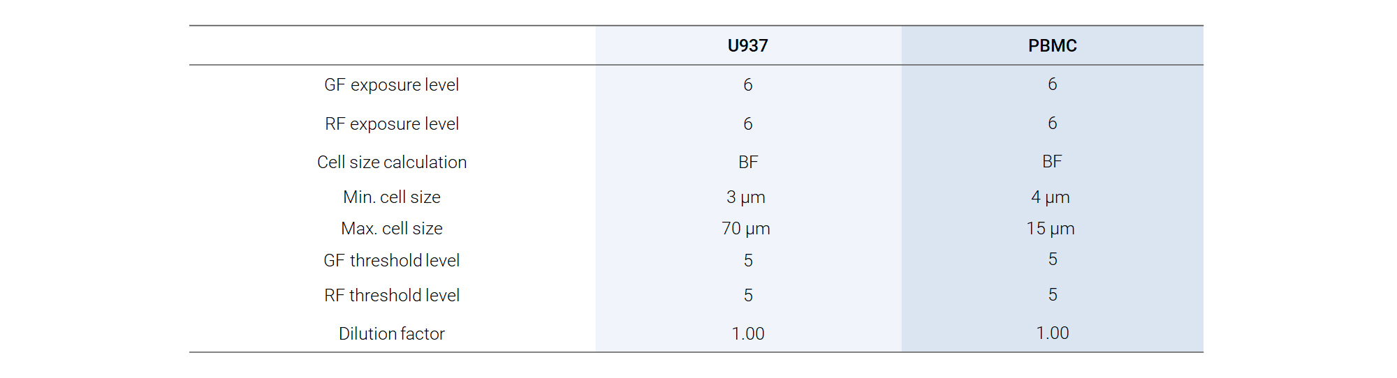
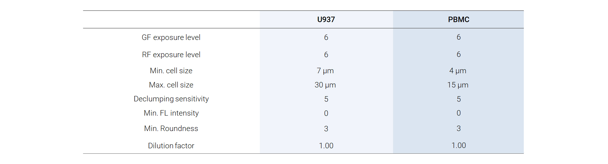
For serial dilution measurements, the cells were diluted with different dilution factors from 1 to 32. A separate batch of cells was prepared for viability measurements with theoretical viability level of 0, 25, 50, 75, and 100%. Cell counts and viability measurements were performed on the LUNA-III™ using the LUNA™ Cell Counting Slide and the built-in PBMC protocol (Table 1). Before loading, the cells were mixed with 0.4% TB (Cat No.T13001) in a 1:1 ratio. The same samples were also used in LUNA-FX7™ to compare the results.
Cell Lines and Reagents
U937 cells and PBMCs were utilized in this study. U937 cells were cultured in RPMI-1640 supplemented with 10 % fetal bovine serum (FBS) and 1 % penicillin/streptomycin. PBMCs were isolated from a 1 mL human peripheral blood sample using the standard density gradient centrifugation technique with Histopaque-1083 (Sigma, #10831). Following final washing steps, the PBMCs enriched in the buffy coat were resuspended in 100 µL of PBS or RPMI medium supplemented with 10 % FBS.
Comparison Between the SpectraSlide® AP-1 and the PhotonSlide™
Cell counting on the LUNA-FX7™ was performed using either the 포톤슬라이드 (PhotonSlide™) (Cat# L12005) or SpectraSlide® AP-1 (Cat# L72061) with modified protocols in Fluorescence Cell Counting mode (Table 1 and Table 2). For 포톤슬라이드 (PhotonSlide™), cells were stained with the standard AO/PI reagent (Cat# F23001) at a 9:1 cell-to-dye ratio, and 10 µL of the stained sample was loaded into the slide chamber. For SpectraSlide® AP-1, 500 µL of cell samples were prepared in 1.5 mL tubes and loaded directly into the slide without additional staining steps.
Linearity of Cell Concentration and Viability
The linearity of cell concentration and viability was evaluated using U937 cells and PBMCs, which were serially diluted from maximum concentrations of 1×10⁷ cells/mL and 5×10⁶ cells/mL, respectively, down to a minimum concentration of 5×10⁵ cells/mL. For viability assessment, live cells were obtained from exponentially growing cultures, while dead cells were generated by heating them at 100 °C for 30 minutes. To create samples with varying viability levels, live and dead cells were mixed at defined ratios of 0, 25, 50, 75, and 100 %. This approach ensured a comprehensive analysis of the system’s ability to accurately differentiate between live and dead cells. All measurements were performed using the LUNA-FX7™ automated cell counter .
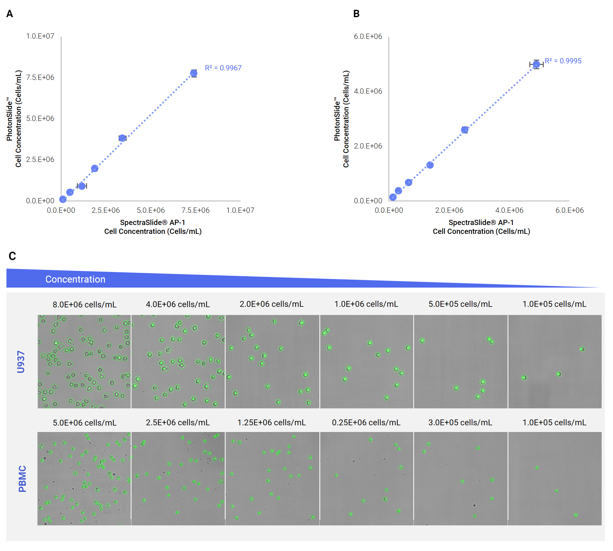
Evaluation of U937 and PBMC Counting Using SpectraSlide® and PhotonSlide™ on the LUNA-FX7™
The performance of the LUNA-FX7™ was assessed by analyzing U937 cells and PBMCs using both the SpectraSlide® and 포톤슬라이드 (PhotonSlide™). The results demonstrated a strong correlation between cell concentrations measured with the two slide types, with R² values of 0.9967 for U937 cells and 0.9995 for PBMCs (Figure 2A and 2B), confirming high linearity and reliability. Additionally, the LUNA-FX7™ accurately identified and classified both U937 and PBMC samples (Figure 2C), highlighting its precision in automated cell counting and reinforcing its effectiveness for consistent and reproducible cell analysis.
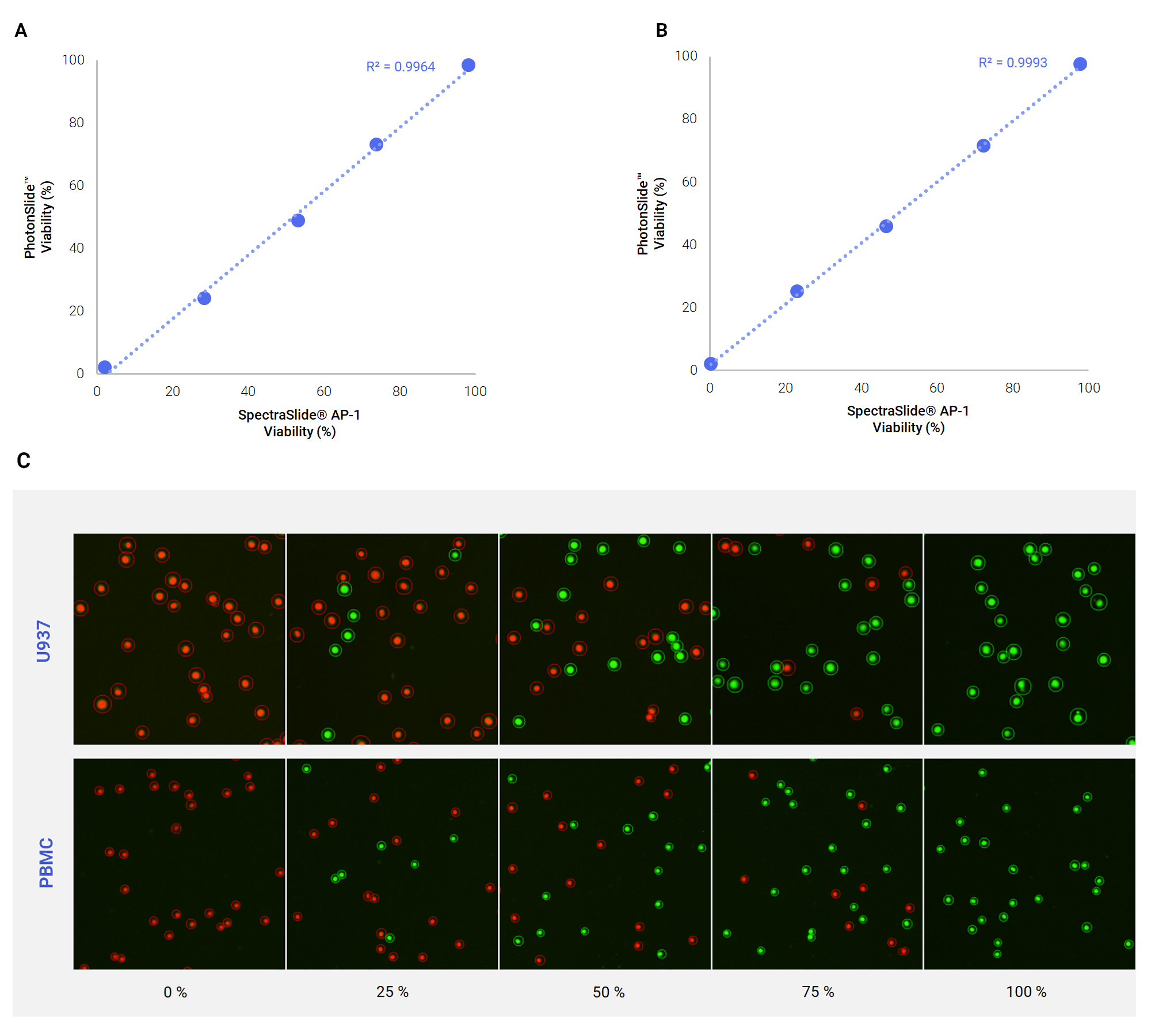
Viability measurements were performed using both the SpectraSlide® and 포톤슬라이드 (PhotonSlide™) and analyzed for comparison. Both slide types exhibited strong correlations with theoretical viability, with R² values of 0.9964 for U937 cells and 0.9993 for PBMCs (Figure 3A and 3B). Viability measurements obtained with SpectraSlide® closely matched PhotonSlide™ results. Across different viability levels, the LUNA-FX7™ showed the great accuracy in live and dead cell tagging when using the SpectraSlide® (Figure 3C), reinforcing its precision in viability assessment.
The SpectraSlide® AP-1 simplifies automated cell counting by integrating sample preparation and staining into a single step. This study validated the strong correlation between SpectraSlide® AP-11 and the widely used 포톤슬라이드 (PhotonSlide™), demonstrating high accuracy in cell concentration (R² = 0.9967 for U937, 0.9995 for PBMCs) and viability assessment (R² = 0.9964 for SpectraSlide®, 0.9993 for PhotonSlide™). Its pipette-free design minimizes handling errors, reduces variability, and enhances workflow efficiency. Offering a reliable, contamination-free solution, the SpectraSlide® AP-1 can provide simple and easy way for cell analysis.