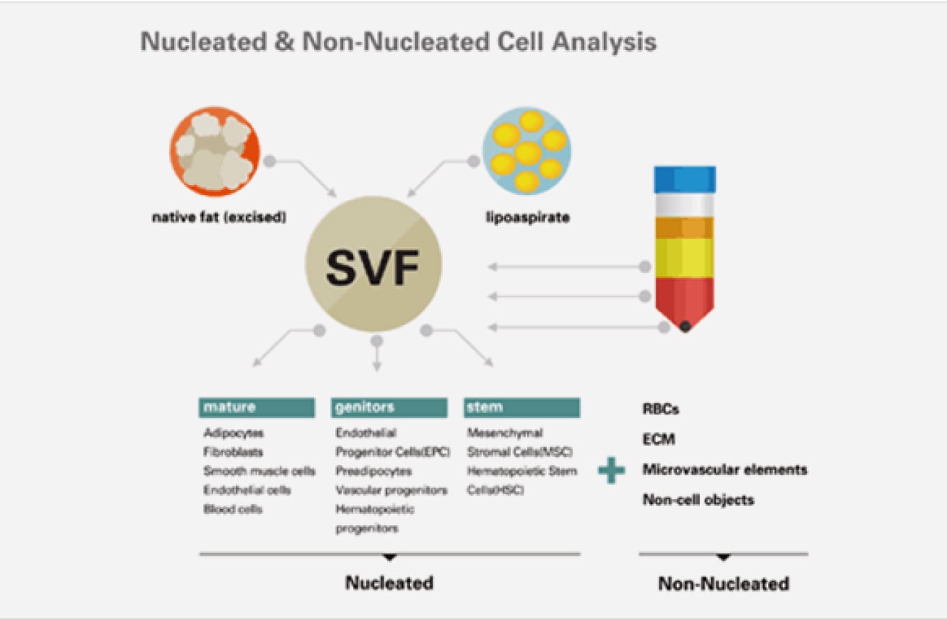
LUNA-STEM™은 지방조직 유래의 SVF 샘플에 함유된 이식 가능한 생존유핵세포의 수를 확인할 목적으로 사용됩니다.
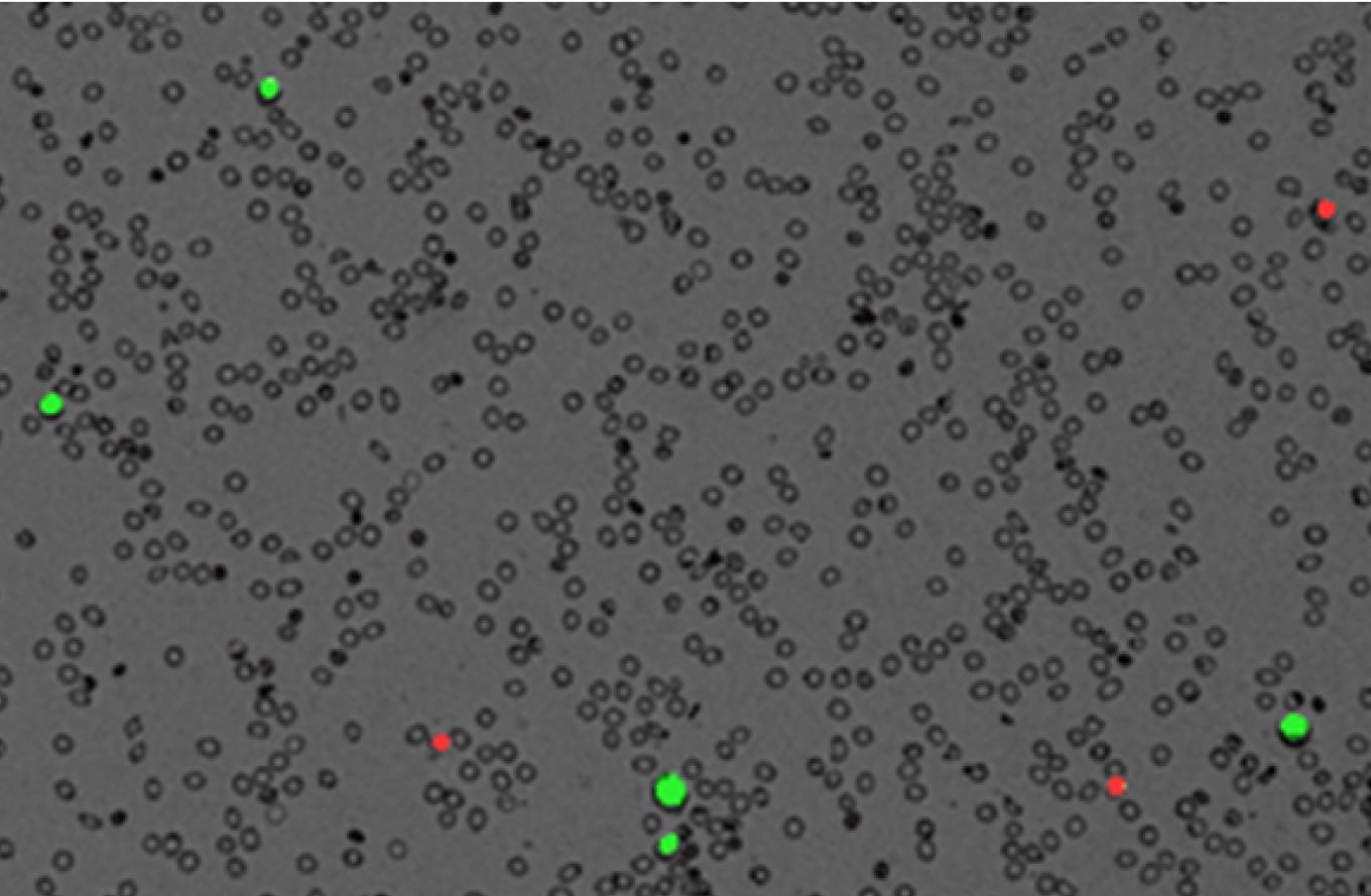
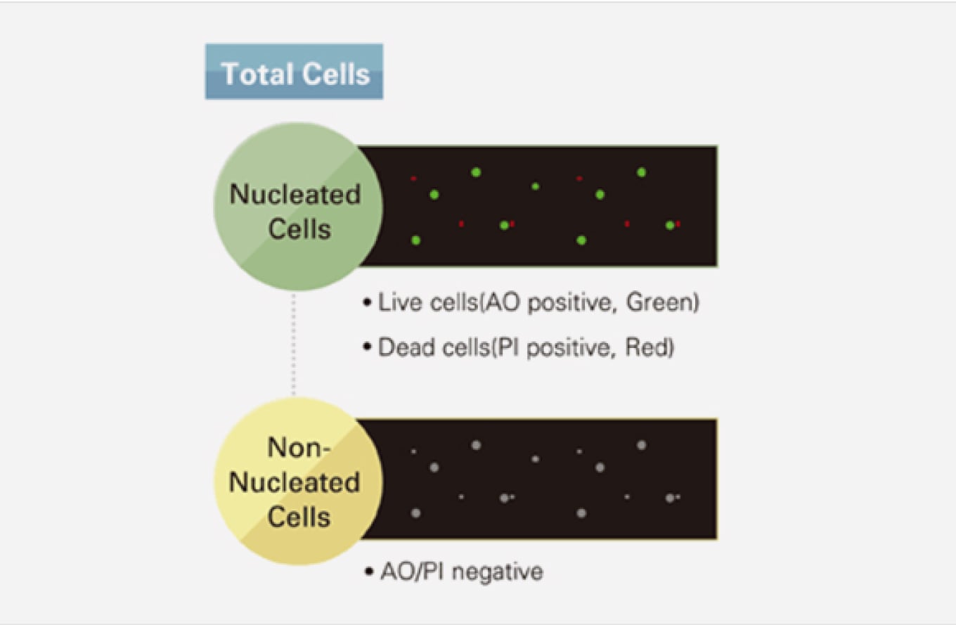
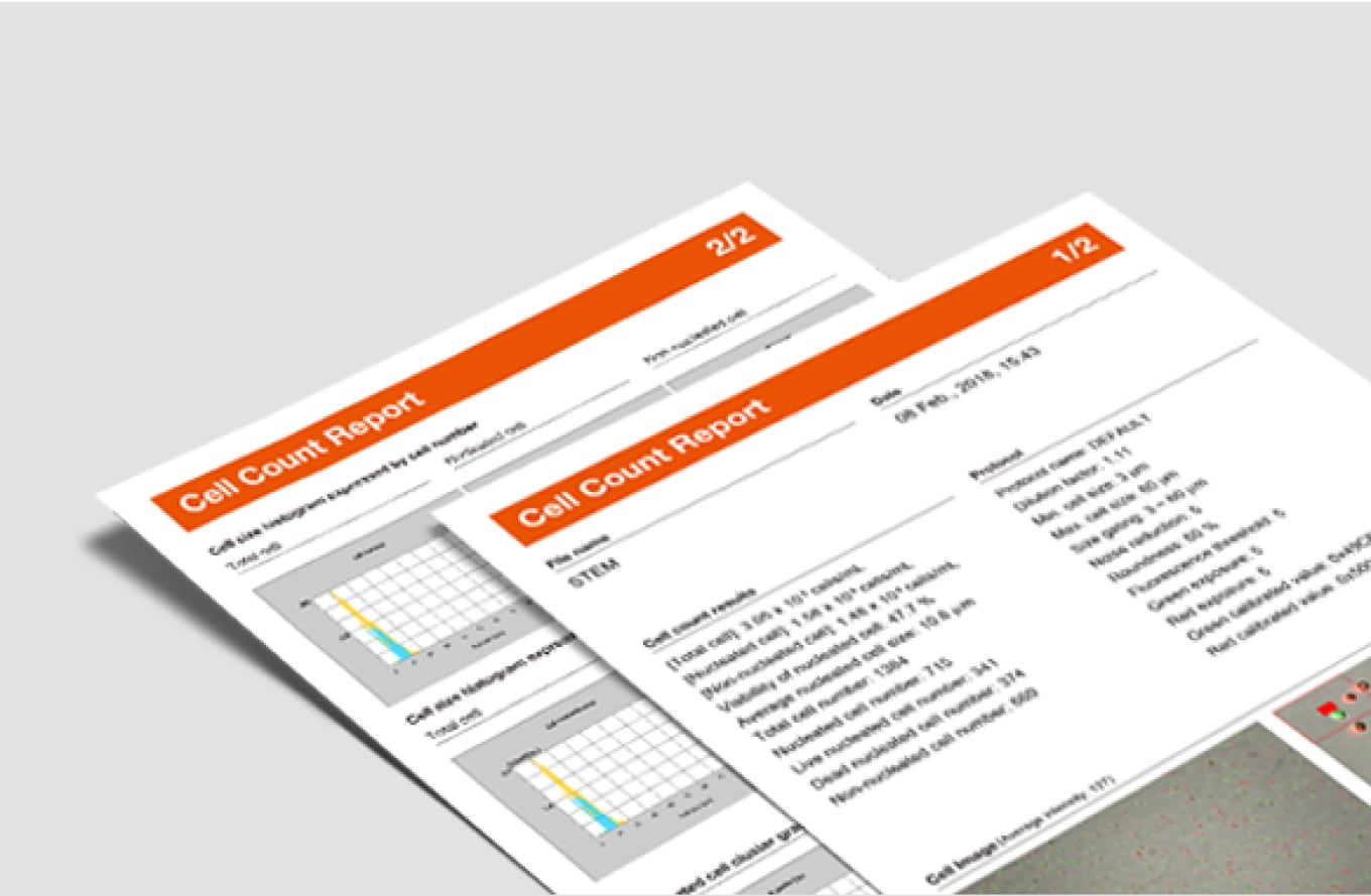
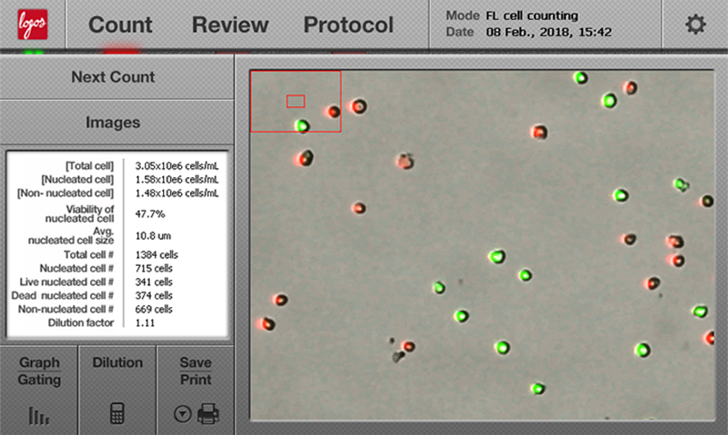
지방조직으로부터 기질혈관분획 (Stromal Vascular Fraction; SVF)을 분리하여 이식세포를 준비할 때, 이식세포의 함량과 생존율은 다음 절차를 진행하기 위해 반드시 확인되어야 할 기본 정보입니다. 일반적으로 조직 유래 샘플에는 적혈구와 세포 파편들도 상당수 존재하므로, 이러한 샘플에서의 세포카운팅은 시간이 오래 걸리고 주관적 오류의 위험도 큽니다. LUNA-STEM™에는 이중형광 광학계와 명시야 광학계가 함께 장착되어 있어 생존유핵세포와 무핵세포를 민감하게 구분할 수 있고, 샘플 내 세포의 핵산을 염색하여 유핵세포의 수와 생존율을 측정할 수 있습니다. 무핵세포 또한 세포파편과 구분되어 카운팅 됩니다. 총 세포수, 유핵세포수, 비핵세포수, 생존율, 세포 크기의 모든 결과를 얻는데 30초면 충분하며, 살아있는 세포는 녹색으로, 죽은 세포는 붉은색으로 표시되는 세포영상에서도 결과를 쉽게 확인할 수 있습니다.

Consistent and accurate day after day!The LUNA-STEM Counter is very quick, effective, and accurate. We use the LUNA-STEM Counter on a daily basis and have run into no troubles for 2 years now. In addition we are a consumer of the LUNA-STEM Slides and AOPI Fluorescent dye, both of which are very inexpensive and extremely obtainable - with help from outstanding customer service. Definitely worth the money.
Best cell counting. Reliable and easy to use.We use it to count mononucleated cells. Very useful and easy to use. Perfect for our daily use.
Hospital de la Santa Creu i Sant Pau
Methods and strategies for procurement, isolation, characterization, and assessment of senescence of human mesenchymal stem cells from adipose tissue.
2019. Gaur M, Dobke M, Lunyak VV. Methods in Molecular Biology.
Serial injections of cryopreserved fat at −196°c for tissue rejuvenation, scar treatment, and volume augmentation.
2018. Ohashi M, Chiba A, Nakai H, Fukuda E, Higuchi T. Plastic and Reconstructive Surgery-Global Open, 6(5): e1742.
Comparison of two automated cell counters for enumeration and viability of mesenchymal stem cells for clinical cellular therapy trials
2018. Radel DJ, Madde P, Dietz A. Cytotherapy, 20(5): S69–S70.
Therapeutic potential of adipose-derived therapeutic factor concentrate for treating critical limb ischemia.
2016. Procházka V, Jurčíková J, Laššák O, Vítková K, Pavliska L, Porubová L, Buszman PP, Krauze A, Fernandez C, Jalůvka F, Špačková I, Lochman I, Jana D, Merfeld-Clauss S, March KL, Traktuev DO, Johnstone BH. Cell transplantation. 25(9): 1623-1633.
The safety and efficacy of cell-assisted fat grafting to traditional fat grafting in the anterior mid-face: an indirect assessment by 3D imaging.
2015. Sasaki GH. Aesthetic Plastic Surgery, 39(6):833-46.
| Cat # | Product | Qty |
|---|---|---|
| L30001 | LUNA-STEM™ Automated Fluorescence Cell Counter for Stem Cells & SVF |
1 unit |
| L12005 | PhotonSlide™, 50 Slides | 1 box |
| L12006 | PhotonSlide™, 500 Slides | 10 boxes |
| L12007 | PhotonSlide™, 1000 Slides | 20 boxes |
| L12011 | LUNA™ Reusable Slide | 1 unit |
| L12012 | LUNA™ Reusable Slides | 2 units |
| L12014 | LUNA™ Reusable Slide Coverslips | 10 units |
| F23001 | Acridine Orange/Propidium Iodide Stain | 2 x 0.5 mL |
| F23002 | Acridine Orange Stain | 2 x 0.5 mL |
| F23003 | Propidium Iodide Stain | 2 x 0.5 mL |
| F23212 | Cell Dilution Buffer | 5 x 20 mL |
| F23102 | LUNA™ Fluorescence Calibration Beads | 1 x 0.5 mL |
| L34003 | LUNA-STEM™ IQ/OQ Protocol | 1 copy |
| P10001 | LUNA™ Printer I | 1 unit |
| P12001 | LUNA™ Printer I Paper (Thermal, 275 prints/roll, 6rolls) | 3 x 2 rolls |
| Sample Volume | 10 μl |
| Cell Counting Time | 30-60 sec (depending on sample conditions) |
| Cell Concentration Range | 5×10⁴ – 1×10⁷ cells/mL (optimal range) |
| Cell Size Range | Detectable Range: 1 – 90 μm Optimal Range: 5 – 60 μm |
| Excitation wavelength | 470 ± 25 nm |
| Emission wavelength | 530 ± 25 nm, 600 nm (LP) |
| Light Source | LED |
| Image Resolution | 5 MP |
| LCD Display | 7 inches (800 x 480 pixels) |
| Dimensions (W×D×H) | 22 × 21 × 9 cm (8.6 × 8.3 × 3.5 in) |
| Weight | 1.8 kg (4 lb) *without the AC adapter |


