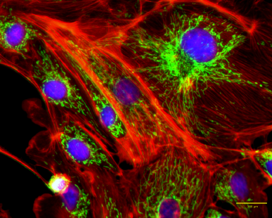
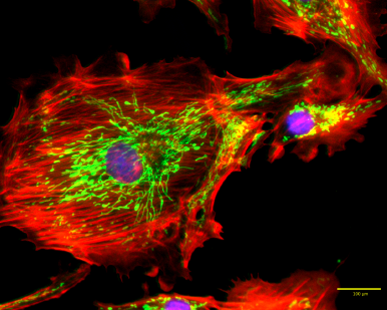
멀티컬러 이미징에서부터 데이터 분석까지
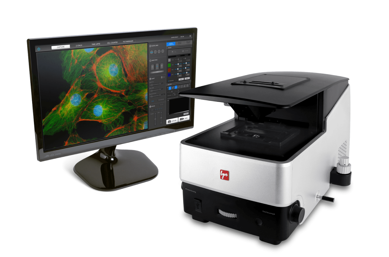
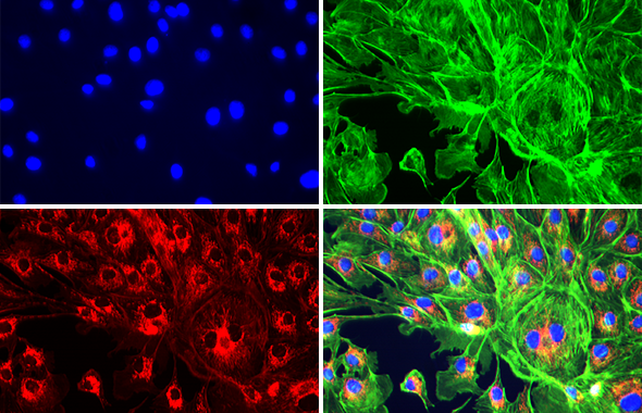
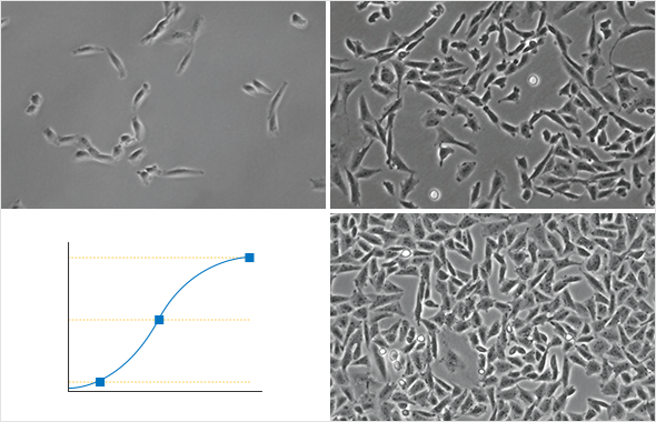
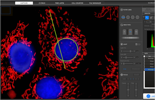
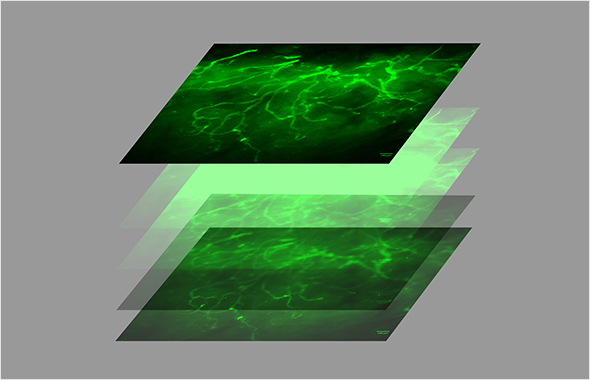
CELENA® S 디지털 이미징 시스템을 사용하면 출판 품질의 고해상도 영상을 손쉽게 촬영할 수 있습니다. 정밀 광학계, 고감도 CMOS 센서, LED 광원 및 형광 필터, 그리고 영상분석 소프트웨어가 설치된 컴퓨터까지 모든 기능들이 CELENA® S 단일 시스템에 통합되어 있습니다. 정교하지만 사용이 편리한 소프트웨어는 다중형광 이미징 (Multicolor Fluorescence Imaging), 명시야 이미징 (BF Imaging), 위상차 이미징 (Phase Contrast Imaging), 타임랩스 라이브 이미징 (Time-Lapse Live Cell Imaging) 및 Z-스택 이미징 (Z-Stack Imaging)을 지원합니다.
CELENA® S에 온스테이지 인큐베이션 시스템을 설치하면 다양한 배양조건에서 라이브 이미징 실험을 수행할 수 있습니다. 가스 혼합기를 함께 사용하면 인큐베이터 내부의 온도, 습도 뿐 아니라 산소함량까지 정밀하게 제어할 수 있습니다. 온도조절기는 가열 장치를 제어하여 인큐베이터 내부의 설정온도를 유지하고 결로를 방지합니다.
또한, 장기배양 중인 세포를 분석할 경우 CELENA® S의 타임랩스 이미징 모드를 이용할 수 있고, 촬영된 영상을 외부로 전송하거나 주석이 첨가된 타임랩스 동영상도 쉽게 제작할 수 있습니다.


It is an essencial for every cell culture lab.
The equipment is very is to use to all its potential, with minimal training. It allows high resolution images of publishing quality.
Dental Medicine, U. Porto
Great equipment. This microscope was excellent value and is simple to use. If you use a vibration isolation table, it is perfectly designed for eliminating the complexities of microscopy without compromise of performance and the Celena makes cell imaging gives optimum results. The Celena imaging system is just right for cell culture.
GPR171 Activation Modulates Nociceptor Functions, Alleviating Pathologic Pain.
2021. Cho PS, Lee HK, Choi YI, Choi SI, Lim JY, Kim M, Kim H, Jung SJ, Hwang SW. Biomedicines 9(3):256.
Rheological properties of cellulose nanofiber hydrogel for high-fidelity 3D printing.
2021. Shin S, Hyun J. Carbohydrate Polymers, 263:117976.
Novel regulatory roles of UCP1 in osteoblasts.
2021. Mukherjee S, Yun JW. Life Sciences 276:119427.
Human WRN is an intrinsic inhibitor of progerin, abnormal splicing product of lamin A.
2021. Kang SM, Yoon MH, Lee SJ, Ahn J, Yi SA, Nam KH, Park S, Woo TG, Cho JH, Lee J, Ha NC, Park BJ. Scientific Reports 11(1):9122.
Inhibition of Lipopolysaccharide-Induced Inflammatory and Oxidative Responses by Trans-cinnamaldehyde in C2C12 Myoblasts.
2021. Park C, Lee H, Hong S, Molagoda IMN, Jeong JW, Jin CY, Kim GY, Choi SH, Hong SH, Choi YH. International Journal of Medical Sciences 18(12):2480-2492.
2022-04-07 | 720 KB
2022-04-07 | 2.14 MB
2021-06-18 | 389.25 KB
2018-04-17 | 526 KB
2018-04-17 | 644 KB
2018-04-17 | 958 KB

| Cat # | Product | Qty |
|---|---|---|
| CS20001 | CELENA® S Digital Imaging System | 1 unit |
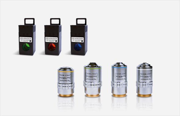
| CS20002 | CELENA® S Digital Imaging System Starter Kit – 4 Objectives – 3 LED Filter Cubes |
1 unit |
| I10520 | CS Stage Top Incubator [Tokai] | 1 set |
| I10501 | Universal Heating System [Ibidi] | 1 set |
| I10502 | Gas Incubation System for CO2 [Ibidi] | 1 set |
| I10503 | Gas Incubation System for CO2/O2 [Ibidi] | 1 set |
| I10201 | Universal Holder | 1 unit |
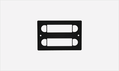
| I10202 | 25 mm x 75 mm Slide Holder, Two Positions | 1 unit |
| I10203 | 35 mm Cell Culture Dish Holder, Four Positions | 1 unit |
| I10204 | 60 mm Cell Culture Dish Holder, Two Positions | 1 unit |
| I10205 | 100 mm Cell Culture Dish Holder, One Position | 1 unit |
| I10206 | 25 c㎡ Nunc T-25 Flask Holder, Two Positions | 1 unit |
| I10207 | 75 c㎡ Nunc T-75 Flask Holder, One Position | 1 unit |
| I10208 | 25 c㎡ BD/Greiner T-25 Flask Holder, Two Positions | 1 unit |
| I10209 | 75 c㎡ BD/Greiner T-75 Flask Holder, One Position | 1 unit |
| I10210 | Glass Hemocytometer Holder, One Position | 1 unit |
| Imaging methods | Epifluorescence and transmitted light (brightfield and phase contrast) |
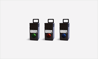
| Illumination | LED filter cubes with adjustable intensity (>50,000 hr life per filter cube) |
| Fluorescence channels | 3 fluorescence channels and 1 transmitted light channel |
| Objective turret | 5 positions |
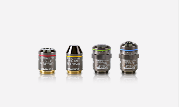
| Objectives | High quality long working distance (LWD) and coverslip-corrected; 1.25X-100X |
| Condenser | 47 mm LWD condenser; 3-positions with brightfield and phase contrast annuli |
| Computer | Built-in dual core CPU, 128 GB SSD internal storage |
| Stage | Mechanical X/Y stage, motorized Z stage; accommodates an onstage incubator |
| LCD display | Full HD color LCD monitor, 1920 x 1080 pixels (not included) |
| Camera | 1.3 MP monochrome CMOS with 1280 x 1024 pixels |
| Images | 8 or 16-bit TIFF, JPG, BMP, or PNG |
| Dimensions (L x W x H) | 44 cm x 30 cm x 27 cm (17.3 x 11.6 x 10.6 in) |
| Weight | 20 kg (44 lb) |


