Discover how the latest automated cell counters are critical to successful single cell genomics workflows.
Automated cell counters have been shown to streamline quality control (QC) of single cell nuclei evaluation, enabling researchers to enhance the success of downstream single cell sequencing workflows.
Accurate quantification of cells is critical for the success of next-generation sequencing (NGS) methods, and some single cell sequencing applications require the isolation of intact nuclei from tissue samples. This has led to increasingly sophisticated and automated cell analysis tools that provide enhanced QC ahead of downstream applications – without which a great deal of time, money and precious sample resource can be wasted in generating sequence information that is misleading or difficult to interpret. The latest automated cell counters, particularly those that utilize fluorescence imaging, provide the necessary QC reassurance, and that is precisely what is required for successful single cell genomics workflows.
Seongwan Jo, an assistant researcher at Logos Biosystems, is engaged in a project using the company’s advanced imaging tools to determine the critical role of accurately evaluating the quality of single nuclei, an important analysis that can help to ensure the success of downstream applications like single cell genomics research. The goal is to identify optimal dye combinations for reliable nuclei assessment across various cell types, tissues and nuclei fixation methods, using Logos Biosystems’ Contador celular automático LUNA-FX7™ and the LUNA-FL™ Contador celular con dos canales fluorescentes.
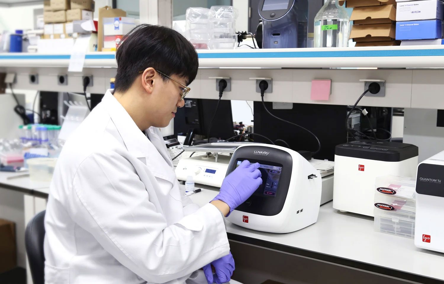
Seongwan Jo, assistant researcher at Logos Biosystems, uses the LUNA-FX7™ Automated Cell Counter for reliable nuclei quantification.
The rise of single cell sequencing
Single cell sequencing enables researchers to study the genomics and transcriptomics of individual cells. With the ability to reveal low-frequency genetic alterations within heterogeneous cell populations that might be missed using bulk sequencing techniques, it has become an important and integral technique to life science research.
“Single cell sequencing enables the analysis of cells characterized by significant heterogeneity through accurately discerning the genetic material of individual cells,” explains Jo. “This contributes to a comprehensive understanding of cellular mechanisms at the genomic level.”
The method allows researchers to characterize abnormal cell populations, determine and analyze rare cells, and map cellular networks. It can also uncover subtle yet important heterogeneities which may have a significant bearing, for example, upon either a patient’s response to a specific therapy or the successful development of an accurate disease model for designing new drugs.
Individual cell and nuclei analyses are crucial for identifying mechanisms that cannot be studied in groups of cells, but their accuracy is entirely dependent upon the quality of the cells and nuclei that are isolated from tissue samples.
The importance of QC in selection of cells and nuclei
Nuclei-based studies have become powerful tools in molecular investigations, particularly with the rise of NGS. “Having high-quality single cell nuclei samples is necessary for single cell sequencing because of its targeted approach, providing advantages in precision, reduced contamination, and a focused examination of nuclear components,” says Jo. He further asserts that nuclei now play a central role in molecular investigations and explains that “nuclei quality assessment has therefore also become a fundamental aspect of genetic studies.”
Downstream sequencing workflows can be seriously compromised by poor nucleic quality. These downstream processes are expensive and time-consuming, and tissue samples are often precious, particularly for example in the case of rare cancers. Wasting such resources through generating misleading sequence information that is difficult to interpret is highly undesirable. The accuracy of final sequence can be negatively affected by poor cell viability, high rates of apoptosis and doublet or multiplet cells. Isolated nuclei can also be misidentified as dead cells.
Reliable, automated cell and nuclei counting methods are therefore a vital first step in establishing successful downstream sequencing workflows and have become critical to avoiding misleading sequence data. They are also now an integral tool in bioprocessing and biopharmaceutical manufacturing, supporting strict regulatory requirements. Not only do they help researchers to determine the molecular properties of an end product, they also make intelligent recommendations about the types of compounds that will help achieve the desired drug.
Fluorescence-based imaging cell counter solutions
A good, automated cell counter must reliably count cells or analyze cell sizes for all sample types, including dissociated tissues, separated nuclei, whole blood and cultured cell lines. Jo describes how the LUNA series of automated cell counters help deliver his research goals: “Utilizing brightfield image data, the LUNA-FX7™ and LUNA-FL™ extract dependable cell size measurements, helping in the evaluation of isolated nuclei quality and mitigating distortions caused by fluorescence intensity,” he explains. According to Jo, this capability is particularly valuable for monitoring size discrepancies before and after nuclei isolation, and he adds that “these automated cell counters quantify the number of nuclei, and fluorescent staining using dyes facilitates the evaluation of cell viability.”
Optimize fluorescent dye combinations
Jo has some useful advice for anyone considering the purchase of the LUNA cell counter series, tailored to specific research needs and objectives. “If your purpose is to assess isolated nuclei, I strongly recommend using fluorescent imaging for better accuracy,” he says. “Moreover, using the appropriate dye combinations is important to ensure successful outcomes. Based on our testing, the Acridine orange (AO)/Propidium Iodide (PI) combination worked the best compared to other dyes,” he further adds.
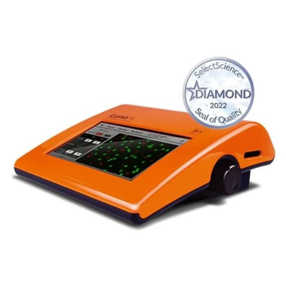
The LUNA-FL™ Dual Fluorescence Cell Counter
Learn more about how the Logos Biosystems’ LUNA-FX7™ and LUNA-FL™ automated cell counters could help you assess nuclei quality for single cell genomics workflows in this application note.


