Keywords
Nuclei isolation is a crucial step in single-cell genomic research, enabling detailed analysis of individual cells from a range of sample types, including fresh tissue, cultured cells, and formalin-fixed paraffin-embedded (FFPE) tissue. It is essential to evaluate nuclei quality following isolation, as it directly influences the accuracy of downstream analyses. Fluorescent-enabled automated cell counters, such as the LUNA-FX7™, simplify this process when combined with fluorescent staining methods. Dual staining with Acridine Orange (AO) and Propidium Iodide (PI) is commonly used for effective nuclei quality assessment.
However, FFPE samples have unique challenges compared to other tissue types due to the formaldehyde fixation and paraffin embedding process. This process compromises nuclei quality, making nuclei isolation more difficult and often resulting in increased debris, which introduces noise and complicates data analysis. To address these challenges, single staining with membrane-impermeable dyes is preferred for FFPE nuclei. This approach reduces noise caused by non-specific dye binding caused by permeable dyes like AO, which tend to bind to debris and interfere with accurate analysis. Nevertheless, we have yet to identify the optimal membrane-impermeable dyes to pair with the LUNA-FX7™ automated cell counter.
In this study, we evaluated membrane-impermeable fluorescent dyes, including PI, NucSpot 470, and YOYO-1, to determine their effectiveness in staining nuclei isolated from kidney FFPE samples using the LUNA-FX7™ automated cell counter. By optimizing dye selection and staining protocols, we aim to improve the reliability of nuclei quality assessments in FFPE tissues, ensuring accurate results and minimizing artifacts caused by debris.
Membrane-impermeable dyes were selected based on their excitation and emission spectra compatibility with the LUNA-FX7™ automated cell counter.
Table 1 . Fluorescent dyes tested for nuclei assessment from FFPE samples.

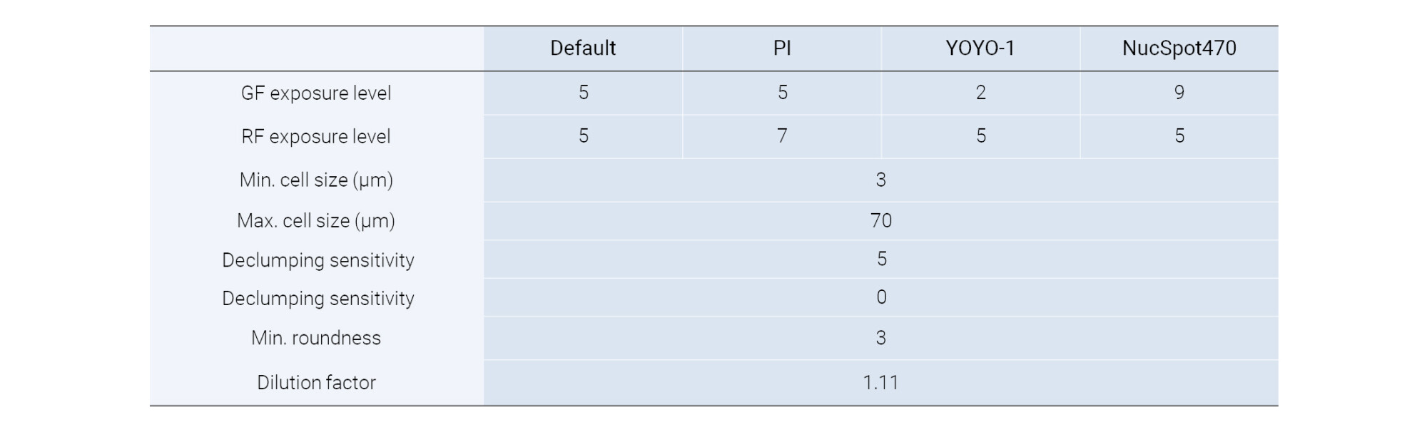
Table 2 . Parameter settings tested for counting nuclei from FFPE samples using the LUNA-FX7™ in Advanced Fluorescence Cell Counting mode.
Staining nuclei from FFPE tissues involves notable challenges due to complex process of isolating nuclei. The process requires melting the paraffin and going through additional isolation steps to access the nuclei. This can compromise nuclei quality and produce more debris compared to fresh samples. This debris may lead to non-specific binding of permeable dyes like AO which compromises staining accuracy; Additionally, the cross-links formed during the fixation process inhibit dye binding to the nuclei, resulting in weaker signals compared to freshly isolated nuclei (Figure 1). Thus, it is important to identify membrane-impermeable dyes that minimize non-specific binding while effectively penetrating fixed nuclei.
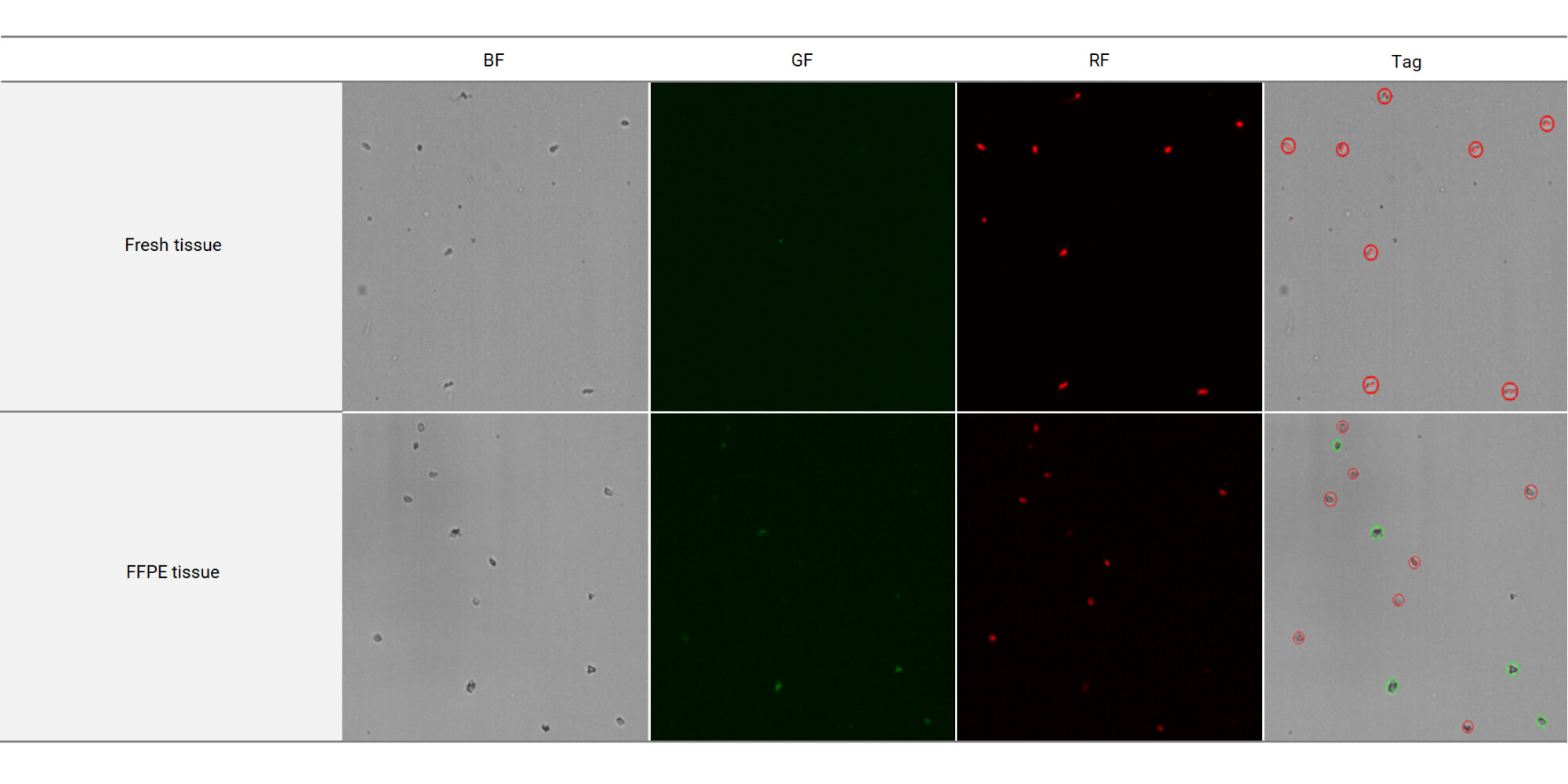
Figure 1 . Comparison of nuclei samples from fresh tissue and FFPE tissue stained using the AO/PI combination, visualized with the LUNA-FX7™ automated cell counter. The top row shows nuclei from fresh tissue and the bottom row represents nuclei from FFPE tissue.
BF: Images captured in the brightfield channel.
GF: Images captured in the green fluorescence channel.
RF: Images captured in the red fluorescence channel.
Tag: Composite images of all channels, fluorescent and brightfield, with identified objects marked using red and green circles. Red circles indicate dead cells,
while green circles represent live cells.
Single impermeable dyes were initially tested using the default protocol of the LUNA-FX7™ automated cell counter, and their performance varied (Figure 2A). PI staining provided a weak signal, although nuclei were tagged; however, increasing the LED level to 7 improved the signal (Figure 2B). YOYO-1 generated a strong signal but introduced slight background noise. Excessive signal strength can lead to the detection of cellular debris, compromising the accuracy of the analysis, but adjusting the GF LED to 2 mitigated this issue (Figure 2C). In contrast, NucSpot470 produced almost no detectable signal, which could not be improved even by increasing the green fluorescence (GF) LED level to 9, demonstrating its limitations when staining isolated nuclei from FFPE samples with the LUNA-FX7™ automated cell counter (Figure 2D).
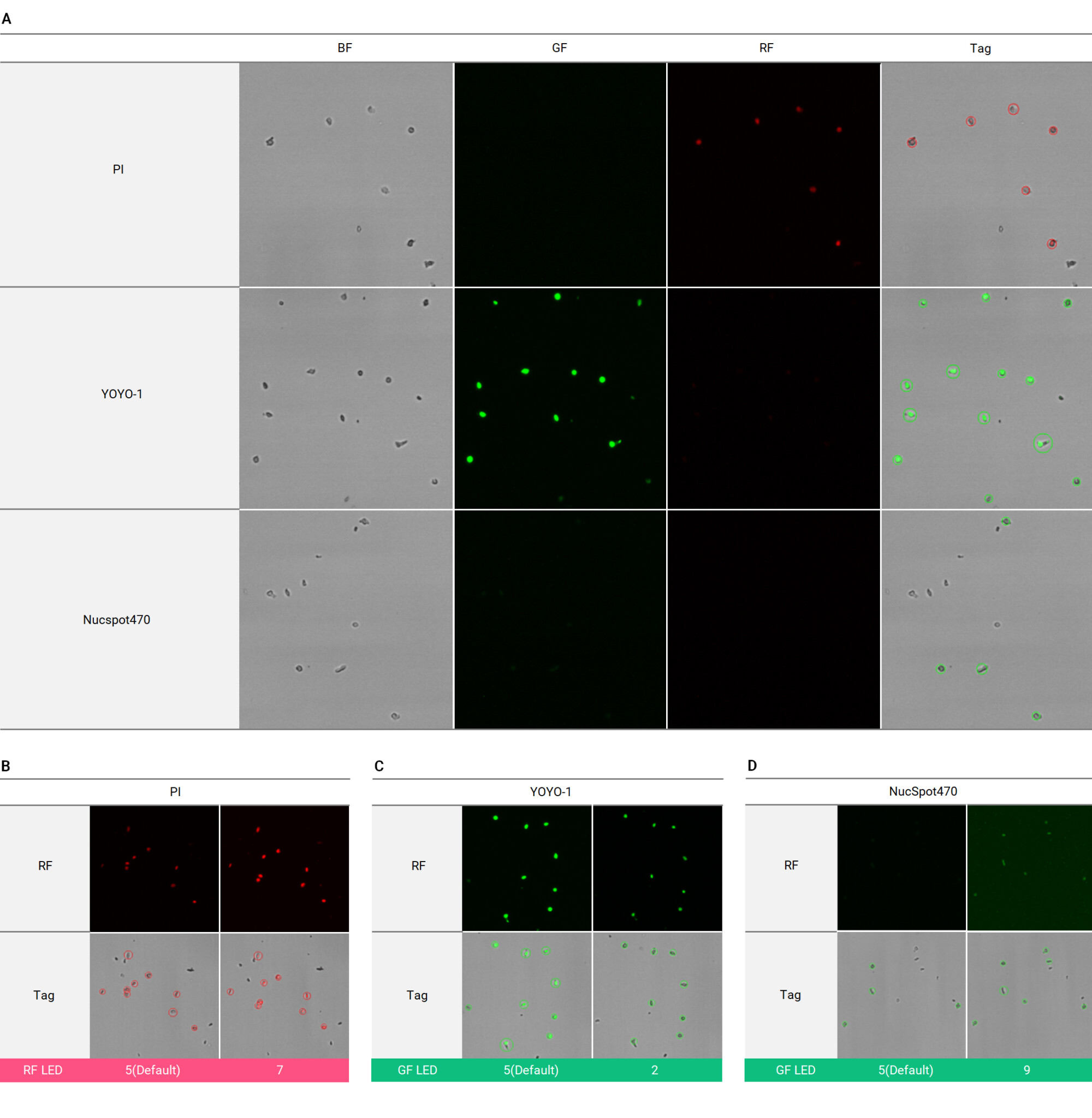
Figure 2 . (A) Evaluation of single impermeable dyes, including PI, YOYO-1, and NucSpot470, for nuclei isolation from FFPE samples using the LUNA-FX7™ automated cell counter. (B) PI staining showed improved signal when the RF LED level was increased from 5 (default) to 7. (C) YOYO-1 displayed strong signal, and reducing the GF LED level from 5 (default) to 2 minimized background noise. (D) NucSpot470 produced minimal signal, even when the GF LED level was increased to 9.
BF: Images captured in the brightfield channel.
GF: Images captured in the green fluorescence channel.
RF: Images captured in the red fluorescence channel.
Tag: Composite images of all channels, fluorescent and brightfield, with identified objects marked using red and green circles. Red circles indicate dead cells, while green circles represent live cells.
In this study, we evaluated the performance of various fluorescent dyes for nuclei assessment in kidney FFPE samples using the LUNA-FX7™ automated cell counter. AO/PI dual staining was found to be ineffective for FFPE samples due to non-specific binding of AO to cellular debris. PI and YOYO-1 showed the best results when LED settings were optimized. PI staining improved significantly when the RF LED level was increased, while YOYO-1 produced strong signals but required adjustments to the GF LED settings to reduce background noise and false signals. In contrast, NucSpot470 demonstrated poor performance even with increased LED settings which indicates its limited suitability for FFPE nuclei assessment. Based on these findings, we recommend using PI or YOYO-1 with optimized LED settings on the LUNA-FX7™ system for reliable nuclei assessment derived from FFPE samples.


