Keywords: LUNA-III™ Automated Cell Counter, Hemocytometer, Cell counting linearity, Cell viability linearity
Accurate cell counting is essential in biomedical research, impacting applications such as cell therapy, disease diagnosis, tissue regeneration, and bioassays. Traditional manual counting methods are time-consuming and prone to variability. Automated systems, like the LUNA-III™ Automated Cell Counter from Logos Biosystems, provide a solution with improved accuracy, reproducibility, and efficiency.
The LUNA-III™ Automated Cell Counter builds on the success of its predecessor, the LUNA-II™, with several key improvements that enhance its functionality and performance. These advancements include live cell detection for large or aggregated cells, new features such as internal storage and better autofocus, the ability to reanalyze data and Find cells feature, and outstanding linearity and low variability in cell concentration and viability measurements.
Introducing LUNA-III™ Automated Cell Counter
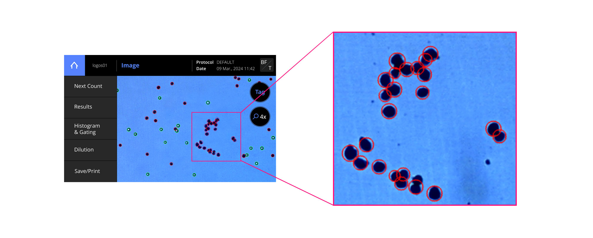
The LUNA-II™ was already a great device, known for its reliability and accuracy in cell counting. However, it had a limitation—declustering of dead cell cluster—that now have been addressed in the LUNA-III™ (Figure 1). Traditional algorithms struggled with declustering dead cells stained with Trypan Blue (TB) due to minimal brightness differences at cell boundaries. LUNA-III™ resolves this with a machine learning algorithm that accurately distinguishes clustered cell shapes. Additionally, LUNA-III™ improves live cell detection by accurately identifying cells with irregular shapes or faint brightness to ensure more reliable results.
In addition to this crucial improvement, the LUNA-III™ boasts several new features designed to enhance user experience and workflow efficiency:
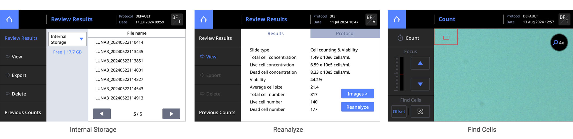
Internal Storage: Save and manage data directly on the device, streamlining your workflow and ensuring easy access to your results.
Reanalyze: Reanalyze previously captured images to ensure accuracy and consistency in your data analysis.
Find Cells: Utilize the ‘Find Cells’ feature to automatically adjust the focal plane and exposure level, ensuring thorough and precise analysis.
These advancements make the LUNA-III™ a superior tool for accurate and efficient cell counting and viability assessment, reinforcing its position as a reliable and innovative solution in the field of cell analysis.
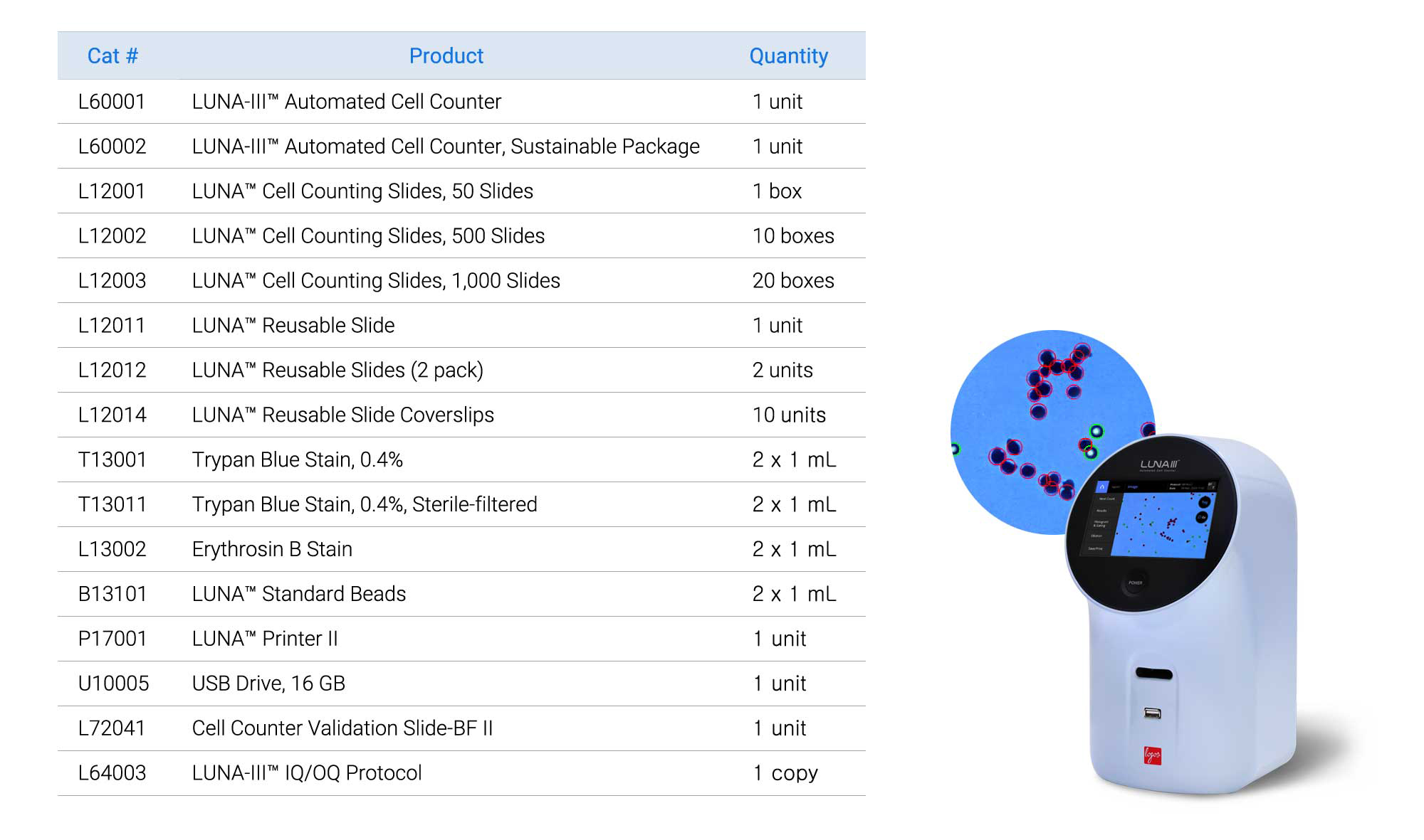
LUNA-III™ Automated Cell Counter
The default protocol of the LUNA-III™ was used for all measurements. Three LUNA-II™ devices were used to test intra-deviation after standard quality control procedures.
Cell Lines and Reagents
U937 (suspension) and 3T3 (adherent) cell lines were used. U937 cells were cultured in RPMI-1640 with 10 % FBS and 1 % penicillin/streptomycin. 3T3 cells were cultured in DMEM with 10 % FBS and 1 % penicillin/streptomycin. 0.4 % trypan blue (Cat No.T13001) was used for viability assessments.
Linearity of Cell Concentration and Viability
Cells were serially diluted starting from a maximum concentration of 1×10⁷ cells/mL (3T3 cells started in maximum concentration 6×10⁶ cells/mL). The cell concentration was measured using the LUNA FX7™ automated cell counter. Live cells were obtained from exponentially growing cultures, while dead cells were prepared by heating cells at 100 °C for 30 min. Mixtures of live and dead cells at varying ratios were prepared to produce samples with different viabilities.
Linearity of Cell Concentration
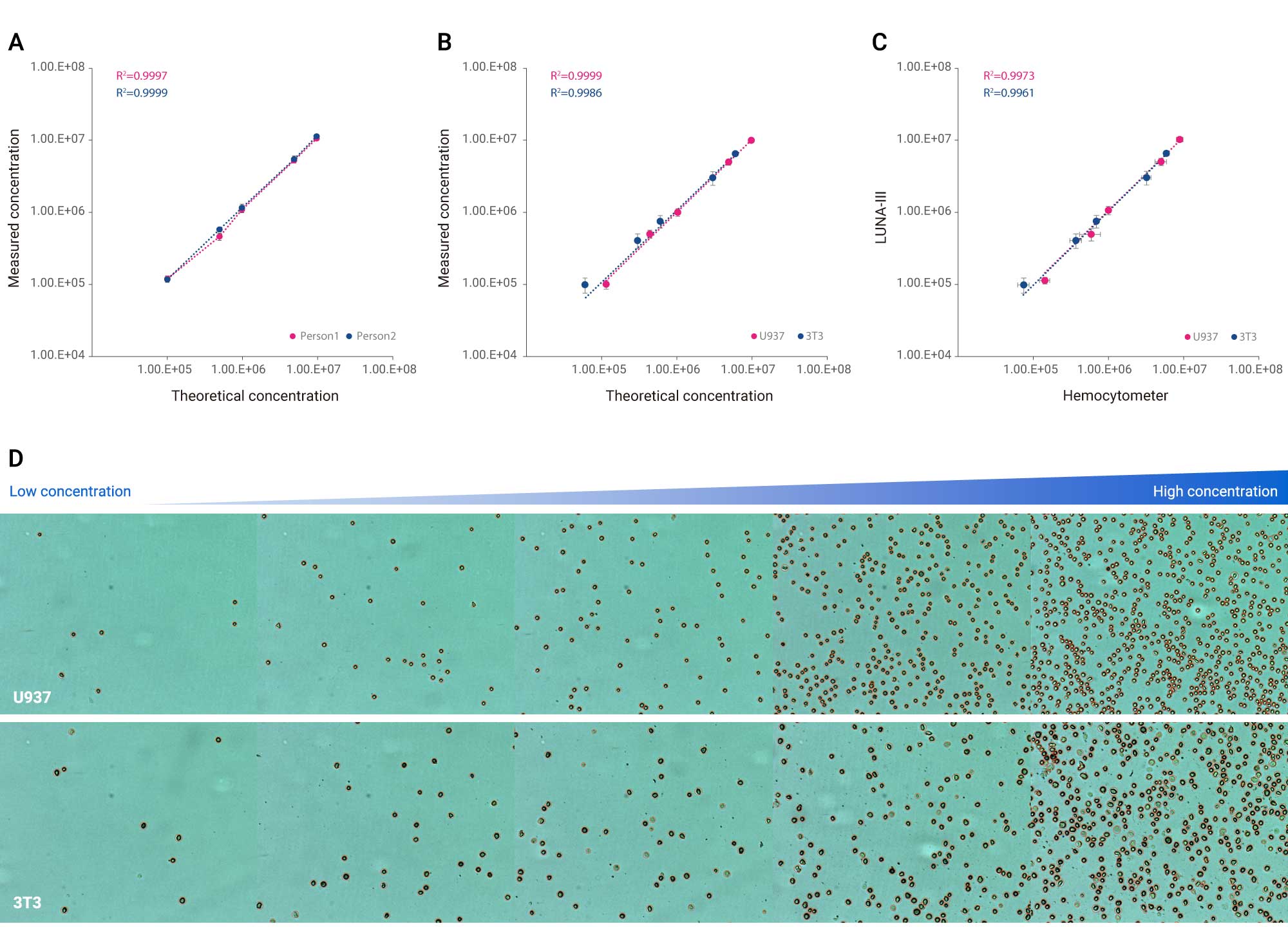
To assess the cell concentration linearity of the LUNA-III™, we conducted tests from three different perspectives: discrepancies between different users (Figure 2A), discrepancies between two cell lines (Figure 2B), and comparison of LUNA-III™ cell counting results with those obtained using a hemocytometer (Figure 2C).
First, we measured linearity using two technicians with varying levels of experience, assessing serially diluted concentrations of U937 cells. Next, we evaluated linearity between U937 and 3T3 cells, which differ in size and characteristics, with a single technician assessing serially diluted samples. Finally, we compared the same samples using a hemocytometer. The LUNA-III™ consistently demonstrated linearity in all scenarios, with R² values of 0.9996 or higher across a broad concentration range. Images show the LUNA-III™ accurately detecting cells in these samples, further illustrating its precision and reliability (Figure 2D). This consistent performance highlights the LUNA-III™’s exceptional ability to accurately measure cell concentrations, showcasing its reliability and precision regardless of user experience.
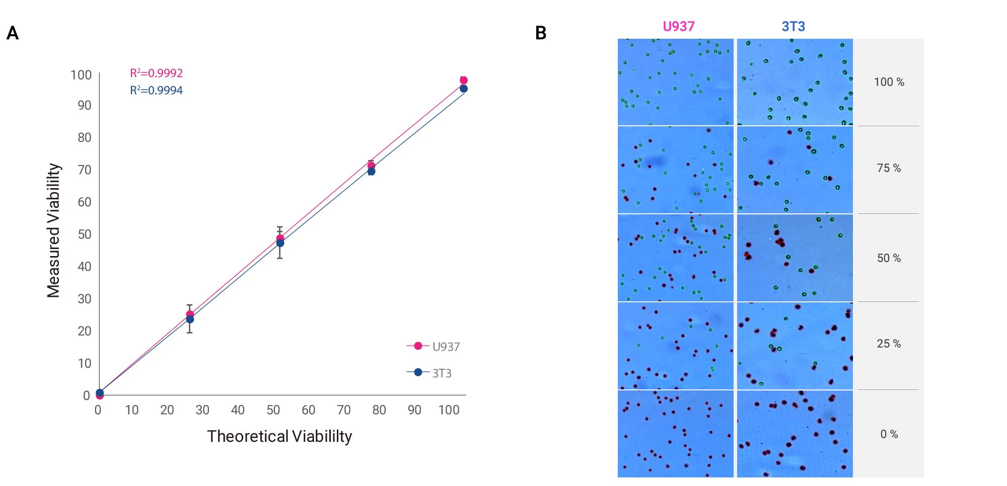
Linearity of Cell Viability
To evaluate the linearity of viability across different cell lines, we utilized the LUNA-III™. Live and dead U937 and 3T3 cells were mixed in varying proportions to create samples with a range of viabilities from 0 % to 100 %, which were then assessed by a single technician. The LUNA-III™ showed exceptional linearity in its viability measurements for both cell lines, achieving R² values of 0.9998 or higher across viability measurements (Figure 3A). The accompanying images show the LUNA-III™’s precise detection of live and dead cells, further highlighting its accuracy and reliability (Figure 3B). This consistent performance underscores the LUNA-III™’s extraordinary capability in measuring cell viability, demonstrating its robust reliability and precision across different cell types.
The LUNA-III™ Automated Cell Counter represents a significant advancement in cell counting and viability assessment, building on the strengths of its predecessor, the LUNA-II™. By addressing key limitations such as cluster detection in dead cells and viability detection in irregularly shaped cells, the LUNA-III™ offers enhanced accuracy and reliability. Its new machine learning algorithms and features like internal storage, reanalysis capability, and the ‘Find Cells’ function make it a powerful tool for researchers. The system’s outstanding linearity in both cell concentration and viability measurements confirms its reliability. Whether for routine cell counting or more complex viability assessments, the LUNA-III™ provides researchers with a reliable, efficient, and user-friendly platform to achieve precise results.