創薬研究および細胞生物学のための自動ハイコンテント画像取得と解析
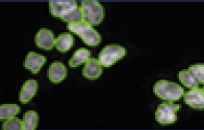
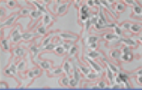
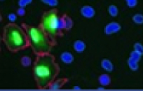
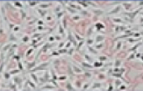
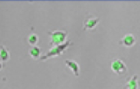
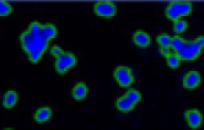
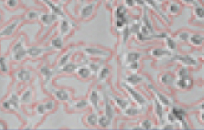
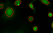
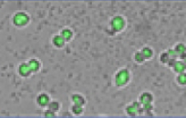
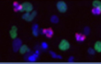
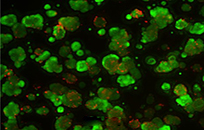
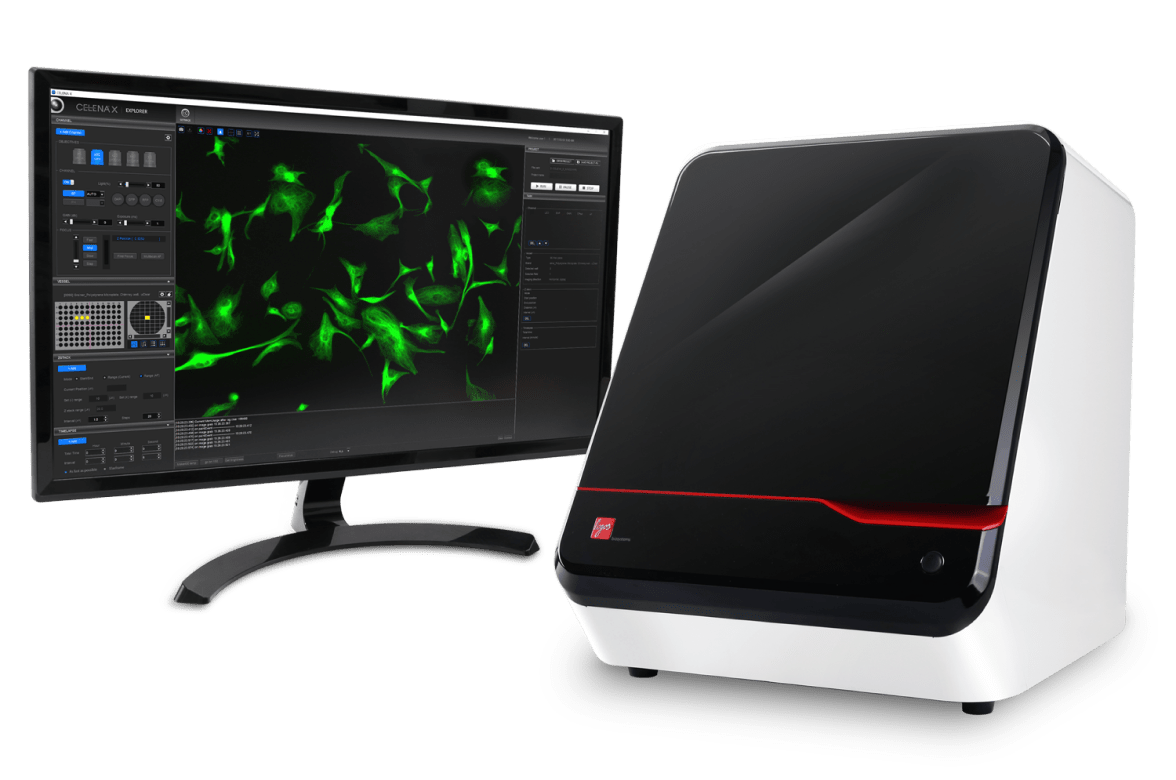
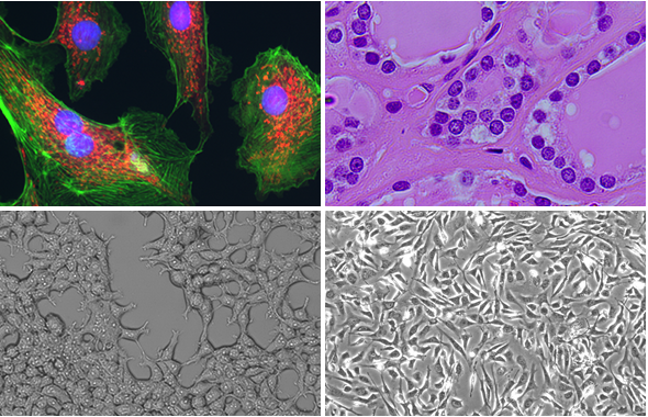
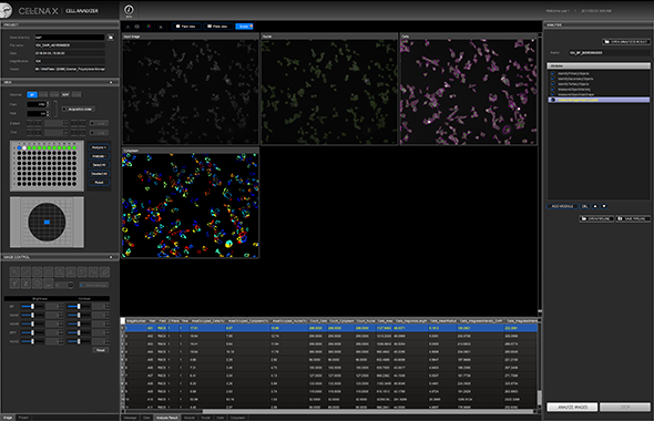
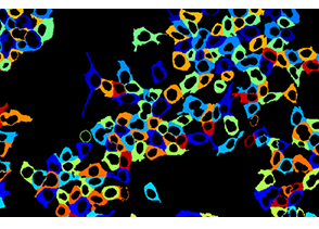
CELENA® X ハイコンテンツイメージングシステムは、迅速な高コンテント画像取得と解析を実現するために設計された統合型イメージングシステムです。カスタマイズ可能な撮像プロトコル、画像ベースおよびレーザーによるオートフォーカス機能、そしてモーター駆動のXYZステージにより、ウェルプレートのイメージングやスライドスキャンが簡単に行えます。 統合されたCELENA® X Cell Analyzer ソフトウェアは、画像とデータを処理し、定量的な解析を可能にします。解析パイプラインは構築・保存が可能で、細胞あるいは細胞内構造の検出、データ収集に最適な画像処理、さまざまな測定の実行などに繰り返し利用できます。 CELENA® Xは、その高性能さと柔軟性を兼ね備えたシステムです。_x000B_交換可能な対物レンズとフィルターキューブにより、固定細胞およびライブセルイメージングの多様なアプリケーションに対応します。


Great to use, inexpensive - watch out big microscopy companies...
The Celena-X is so easy to use, it has huge flexibility and it is oh so affordable making the 'big name brands' look like a rip off!
ATA Scientific
Perfect combination of high content and high quality imaging.
The speed of image acquisition is outstanding and the images generated are of high quality. Together with the easy-to-use software, it is a great product.
Institut für Zellbiologie und Immunologie Stuttgart
Primary cilia and the reciprocal activation of AKT and SMAD2/3 regulate stretch-induced autophagy in trabecular meshwork cells.
2021. Shim MS, Nettesheim A, Dixon A, Liton PB. Proceedings of the National Academy of Sciences of the United States of America 118(13):e2021942118.
Cathepsin B Localizes in the Caveolae and Participates in the Proteolytic Cascade in Trabecular Meshwork Cells. Potential New Drug Target for the Treatment of Glaucoma.
2021. Nettesheim A, Shim MS, Dixon A, Raychaudhuri U, Gong H, Liton PB. Journal of Clinical Medicine 10(1):78.
Indirubin-3'-monoxime induces paraptosis in MDA-MB-231 breast cancer cells by transmitting Ca 2+ from endoplasmic reticulum to mitochondria.
2021. Dilshara MG, Neelaka Molagoda IM, Prasad Tharanga Jayasooriya RG, Choi YH, Park C, Kim GY. Archives of Biochemistry and Biophysics 698:108723.
Revascularization and limb salvage following critical limb ischemia by nanoceria-induced Ref-1/APE1-dependent angiogenesis.
2020. Park IS, Mahapatra C, Park JS, Dashnyam K, Kim JW, Ahn JC, Chung PS, Yoon DS, Mandakhbayar N, Singh RK, Lee JH, Leong KW, Kim HW. Biomaterials 242:119919.
Synthesis and Properties of CurNQ for the Theranostic Application in Ovarian Cancer Intervention.
2020. Freidus LG, Kumar P, Marimuthu T, Pradeep P, Pillay V, Choonara YE. Molecules (Basel, Switzerland) 25(19):4471.
2022-04-07 | 2 MB
2021-07-01 | 282.34 KB
2021-06-18 | 665.56 KB
2019-10-30 | 901.13 KB
2023-08-16 | 132 KB
2022-06-07 | 664.37 KB
2022-05-10 | 254.01 KB
2022-04-08 | 399.60 KB
2022-02-09 | 141.67 KB
2021-12-15 | 378.75 KB
2021-10-19 | 3.61 MB
2021-09-07 | 455.78 KB
2019-05-28 | 456.95 KB
2019-03-25 | 422.40 KB
2019-03-07 | 356.63 KB
2019-01-16 | 574.17 KB
2018-12-07 | 417.63 KB
| Cat # | Product | Qty |
|---|---|---|
| CX30000 | CELENA® X High Content Imaging System | 1 unit |
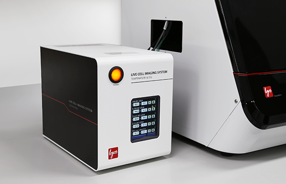
| I10530 | CX Stage Top Incubator, Basic [Tokai] | 1 set |
| I10531 | CX Stage Top Incubator, Pro [Tokai] | 1 set |
| I10500 | Heating System for Multi-well Plates [Ibidi] | 1 set |
| I10502 | Gas Incubation System for CO2 [Ibidi] | 1 set |
| I10503 | Gas Incubation System for CO2/O2 [Ibidi] | 1 set |
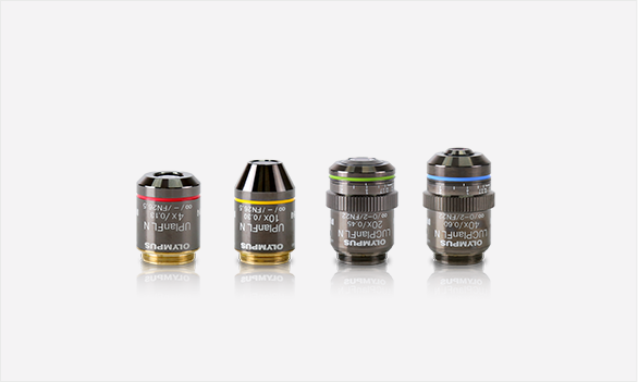
| I10030 | UPLFLN 4X, Olympus | 1 unit |
| I10031 | UPLFLN 10X2, Olympus | 1 unit |
| I10034 | LUCPLFLN 20X, Olympus | 1 unit |
| I10035 | LUCPLFLN 40X, Olympus | 1 unit |
| I10038 | UPLFLN 4XPH, Olympus | 1 unit |
| I10039 | UPLFLN 10X2PH, Olympus | 1 unit |
| I10042 | LUCPLFLN 20XPH, Olympus | 1 unit |
| I10043 | LUCPLFLN 40XPH, Olympus | 1 unit |
| I10046 | PLAPON 1.25X, Olympus | 1 unit |
| I10047 | PLAPON 2X, Olympus | 1 unit |
| I10052 | UPLXAPO 60XO, Olympus | 1 unit |
| I10051 | UPLXAPO 100XO, Olympus | 1 unit |
| I10130 | DAPI (Ex395/25, Em460/50) | 1 unit |
| I10131 | EGFP (Ex470/30, Em530/50) | 1 unit |
| I10132 | RFP (Ex530/40, Em605/55) | 1 unit |
| I10133 | mCherry (Ex580/25, Em645/75) | 1 unit |
| I10134 | ECFP (Ex436/20, Em480/40) | 1 unit |
| I10135 | EYFP (Ex500/20, Em535/30) | 1 unit |
| I10136 | DSRed (Ex530/40, Em620/60) | 1 unit |
| I10137 | Cy5 (Ex620/60, Em700/75) | 1 unit |
| I10138 | Cy7 (Ex710/75, Em810/90) | 1 unit |
| I10139 | Cy3/TRITC Long Pass (Ex530/40, Em570lp) | 1 unit |
| I10140 | GFP Long Pass (Ex470/40, Em500lp) | 1 unit |
| I10141 | Cy5 Long Pass (Ex620/60, Em665lp) | 1 unit |
| I10142 | Custom Filters | 1 unit |
| I10410 | Joystick | 1 unit |
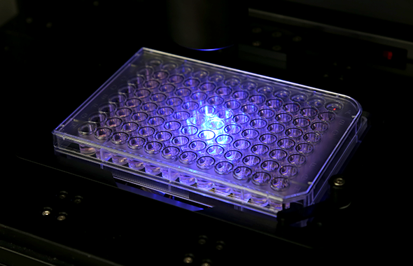
| Supported labware | Slides, multi-well plates (6 to 1536 wells), petri dishes, culture flasks |
| Imaging modes | 4-channel fluorescence, brightfield, phase contrast, color brightfield |
| Light source | High-power LED filter cubes with adjustable intensity (>50,000 hours per filter cube) |
| Filter cube stage | Motorized; 4 interchangeable fluorescence filter cubes and 1 brightfield filter cube |
| Available filters | DAPI, EGFP, RFP, mCherry, ECFP, EYFP, DSRed, Cy5, Cy7, Cy3/TRITC Long Pass, GFP Long Pass, Cy5 Long Pass, custom filters |
| Objective turret | Motorized; 5 interchangeable objectives |
| Compatible objectives | 1.25-100X; Olympus, Zeiss, and Logos Biosystems objectives |
| Condenser | Motorized; basic or phase contrast Basic: 60 mm LWD condenser, 4 positions Phase contrast: 60 mm LWD condenser, 4 positions with 3 phase annuli |
| Camera | Monochrome: CMOS, 1.92 MP (optional) Color: CMOS, 1.92 MP |
| Image outputs | Monochrome: 16‐bit (12‐bit dynamic range) TIF, PNG, or JPG Color: 24-bit color TIF, PNG, or JPG Movies: MP4 |
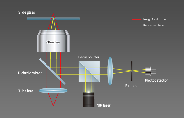
| Autofocus method | Image-based autofocus (optional) Laser autofocus |
| Stage | Motorized X/Y-stage (120 mm x 80 mm); motorized Z-stage (10 mm) |
| Stage control | CELENA® X Explorer (optional) Joystick |
| Computer | External PC |
| Monitor | 27” 4K UHD monitor |
| Software | User interface: CELENA® X Explorer Analysis: CELENA® X Cell Analyzer |
| Power | 100-240 VAC, 250 W, 50/60 Hz |
| Dimensions | Main body: 39 x 46 x 50 cm (15.4 x 18.1 x 19.7 in) Controller: 17 x 30 x 23 cm (6.7 x 11.8 x 9.1 in) |
| Weight | Main body: 33 kg (72.8 lbs) Controller: 7 kg (15.4 lbs) |