Keywords
Mammalian cell lines such as CHO-K1 and HEK293 are essential for the production of therapeutic proteins, monoclonal antibodies, and viral vectors, and are widely utilized in biopharmaceutical research and development. These cell lines are preferred for their robust protein expression, human-compatible post-translational modifications, and suitability for both transient and stable expression systems. As protein production is closely linked to the health and density of the culture, precise and reliable measurement of cell concentration and viability is critical for optimizing transfection efficiency and maximizing protein yield.
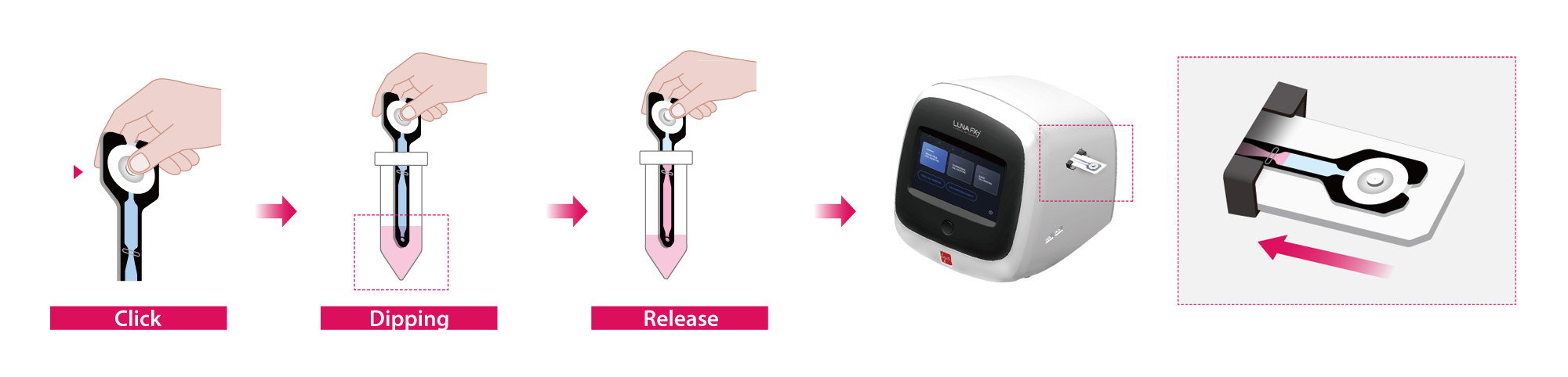
The LUNA-FX7™ Automated Cell Counter, in combination with Acridine Orange (AO) and Propidium Iodide (PI) dual fluorescence staining, provides a fast and reliable platform for assessing cell viability in mammalian cultures. Traditionally, the PhotonSlide™ has been used as the standard for sample loading in these assays. However, the newly developed SpectraSlide® AP-1 offers a significant enhancement to this workflow. Featuring a pre-coated
AO/PI surface and an intuitive “click–dip–release” operation, the SpectraSlide® AP-1 eliminates the need for separate dye preparation and manual sample loading. This innovation not only reduces hands-on time but also enhances assay consistency, workflow efficiency, and reproducibility.
In this application note, we evaluate the performance of the SpectraSlide® AP-1 for viability and concentration analysis of CHO-K1 and HEK293 cells using the LUNA-FX7™, and compare its results to those obtained with the PhotonSlide™.
Cell Culture
CHO-K1 cells were cultured in RPMI-1640 medium, and HEK293 cells were maintained in DMEM. Both media were supplemented with standard additives, including fetal bovine serum (FBS), antibiotics, or other necessary growth factors, following standard cell culture practices.
Cell Counting and Analysis
Cell counting was conducted on the LUNA-FX7™ automated cell counter using either the PhotonSlide™ (Cat# L12005) or SpectraSlide® AP-1 (Cat# L72061) by using the DEFAULT protocol. For PhotonSlide™, the 2 Ch mode was selected, and 18 µL of the cell mixture was stained with Acridine Orange/Propidium Iodide (AO/PI) reagent (Cat# F23001) at a 9:1 ratio. 10 µL of the stained sample was loaded into the slide chamber. For SpectraSlide® AP-1, the Spectra mode was selected. 500 µL of cell samples were prepared in 1.5 mL tubes and loaded directly into the slide without additional staining steps.
RESULTS
To evaluate the performance of the SpectraSlide® AP-1 in cell counting and viability assessment, we compared it to the PhotonSlide™ using CHO-K1 and HEK293 cells.
Representative montage images (Figure 2 A and 2 B) clearly demonstrated the ability of both slide types to differentiate live and dead cells. On both the PhotonSlide™ and SpectraSlide® AP-1, live cells stained with AO appeared green, while dead cells stained with PI appeared red. The images revealed strong signal contrast and accurate segmentation, with no observable dye precipitation or background interference on either slide type.
Cell concentration values obtained from the SpectraSlide® AP-1 closely matched those measured with the PhotonSlide™, as shown in CHO-K1 and HEK293 cells (Figure 2 C and 2 D). For CHO-K1 cells, total cell counts averaged around 2.3 × 10⁶ cells/mL, while HEK293 cells showed slightly lower values near 2.2 × 10⁶ cells/mL. The minimal variation between the two slide types demonstrates the reproducibility and counting accuracy of the SpectraSlide® AP-1. Viability assessments also showed high consistency across both slides (Figure 2 E and 2 F). Samples intentionally prepared at ~50 % viability yielded nearly identical results regardless of the slide used.
Figure 2. (A, B) Representative montage images of CHO-K1 (A) and (B) cells captured on the LUNA-FX7™. The left panel shows unstained brightfield images, while the right panel displays fluorescence overlays with viable cells stained green and non-viable cells stained red.
(C, D) Bar graphs comparing total cell concentrations obtained using the PhotonSlide™ and SpectraSlide® AP-1 for CHO-K1 (C) and HEK293 (D) cells.
(E, F) Viability percentages for CHO-K1 (E) and HEK293 (F) cells, showing close agreement between the two slide types.
BF: Images captured in the brightfield channel.
Tag: Composite images of all channels, fluorescent and brightfield, with identified objects marked using red and green circles. Red circles indicate dead cells, while green circles represent live cells.
CONCLUSION
This study shows that the SpectraSlide® AP-1 is a reliable and easy-to-use tool for measuring cell concentration and viability when used with the LUNA-FX7™ Automated Cell Counter. Its performance was very similar to the well-known PhotonSlide™, proving that it works well with both CHO-K1 and HEK293 cells. The built-in AO/PI staining and simple “click–dip–release” workflow help reduce hands-on time and limit user error. With clear imaging of live and dead cells, the SpectraSlide® AP-1 is a strong choice for fast, consistent cell analysis in everyday biopharmaceutical research and production.


