What Is Cell Death? Experimental Distinction Between Apoptosis, Necrosis, and Necroptosis
2026-01-05
What Is Cell Death? Experimental Distinction Between Apoptosis, Necrosis, and Necroptosis
Cell death is a fundamental biological process essential for understanding life phenomena, disease mechanisms of action (MOA), and cellular responses to drugs. To ensure the reliability of experimental results, it is not sufficient to simply conclude that “cells have died”; rather, it is critical to accurately identify which pathway of cell death has occurred.
In this post, we introduce the three most important forms of cell death—apoptosis, necrosis, and necroptosis—collectively referred to here as the “Big Three of Cell Death.” We provide an in-depth comparison of these pathways from both morphological and biochemical perspectives, with a particular focus on how they can be clearly distinguished using experimental approaches in the laboratory.
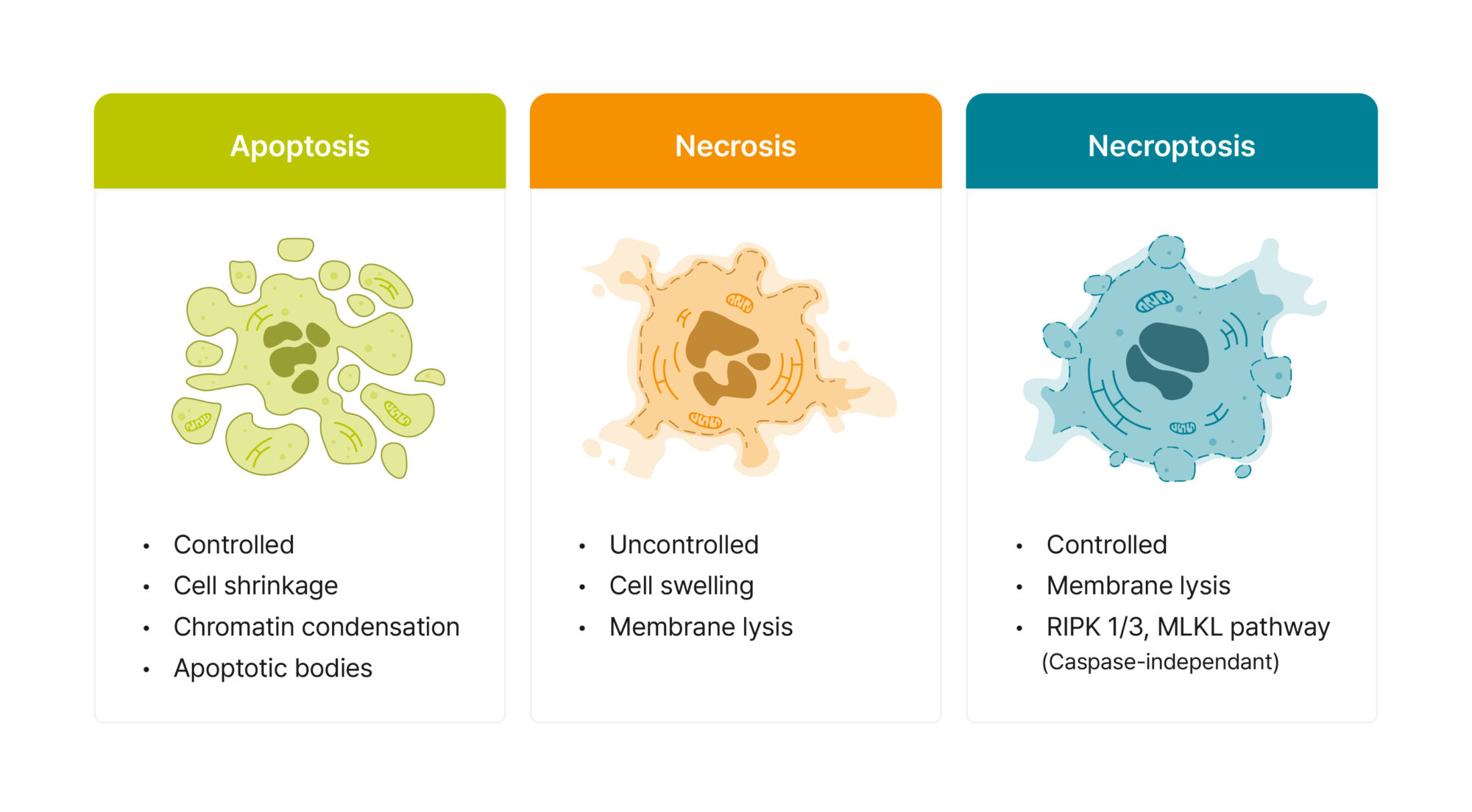
Figure 1. Morphological characteristics of the three major types of cell death
1. Programmed Cell Death: Apoptosis
Apoptosis is a form of programmed cell death in which cells actively and tightly regulate their own demise. This process is characterized by minimal damage to the surrounding environment and is often described as a relatively “silent” mode of cell elimination.
Morphological features
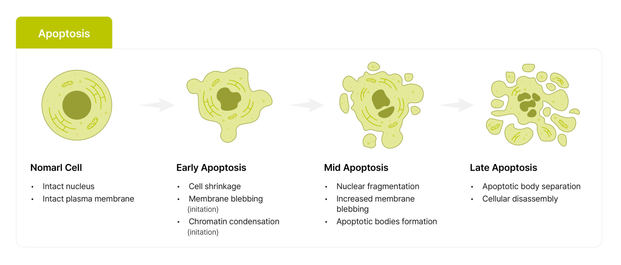
Cell shrinkage
Membrane blebbing accompanied by the formation of apoptotic bodies
Chromatin condensation and nuclear fragmentation
Preservation of plasma membrane integrity (at early stages)
Biochemical features
Caspase activation, with executioner caspases such as caspase-3 and caspase-7 playing central roles
DNA laddering, in which genomic DNA is cleaved into regular fragments of approximately 180–200 bp
Externalization of phosphatidylserine (PS), a membrane phospholipid that becomes exposed on the outer leaflet of the plasma membrane
ATP-dependent signaling and processes (which may vary depending on cellular context)
Major triggers
Elimination of unnecessary cells during development
DNA damage
Growth factor deprivation
Attack by cytotoxic T lymphocytes
Inflammatory response
During apoptosis, cells are fragmented into apoptotic bodies, which are rapidly recognized and cleared by macrophages through “eat-me” signals (such as PS exposure). As a result, cellular contents are efficiently contained and do not leak into the extracellular space, minimizing inflammatory stimulation of surrounding tissues.
However, in in vitro conditions where phagocytic clearance is insufficient, or when apoptotic cells are left uncleared for extended periods, apoptosis may progress to secondary necrosis. In this case, plasma membrane integrity is eventually lost, intracellular contents are released, and inflammatory signals similar to those observed in necrosis can be detected. Therefore, in experimental settings, it is safer to interpret apoptosis by considering both the time course and the efficiency of cell clearance, rather than assuming that apoptosis is always non-inflammatory.
Figure 2. Stage-dependent morphological changes during apoptosis
2. Unregulated Cell Death: Necrosis
Necrosis traditionally refers to a form of cell death in which cells are uncontrollably destroyed as a result of severe insults such as toxins, physical trauma, or extreme stress.
Morphological features
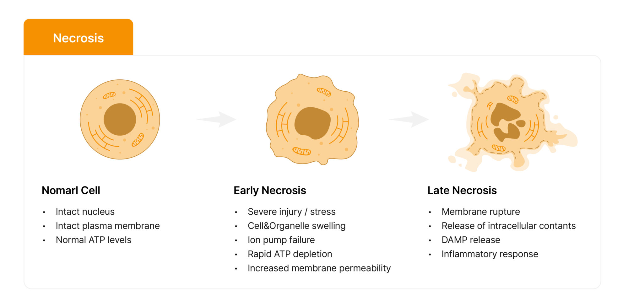
Cell and organelle swelling
Plasma membrane rupture (lysis) and release of intracellular contents
Karyolysis, characterized by nuclear dissolution without chromatin condensation
Biochemical features
Inactivation of caspases
Rapid depletion of ATP, leading to failure of ion pump function
Random DNA fragmentation, observed as a smear pattern on gel electrophoresis
Increased plasma membrane permeability
Major triggers
Physical trauma
Exposure to toxic substances
Ischemia / hypoxia
Extreme temperature changes
Metabolic dysfunction
Inflammatory response
Necrosis is frequently associated with a strong inflammatory response, as rapid disruption of the plasma membrane leads to the release of intracellular components into the extracellular space. These components, known as damage-associated molecular patterns (DAMPs), activate surrounding immune cells and promote inflammation.
Importantly, the intensity of inflammation is often more closely related to when and how plasma membrane integrity is lost—that is, how lytic the terminal stage of cell death is—rather than to the specific name assigned to the cell death pathway itself.
Figure 3. Stage-dependent morphological changes during necrosis
3. Programmed Necrosis: Necroptosis
Necroptosis is a form of programmed necrosis that, from a morphological standpoint, resembles necrosis in that cells undergo swelling, loss of plasma membrane integrity, and release of intracellular contents, ultimately exhibiting a lytic mode of cell death. However, unlike accidental necrosis, necroptosis is tightly regulated by specific intracellular signaling pathways.
Necroptosis is particularly notable in conditions where the apoptotic pathway is suppressed—such as during caspase-8 inhibition or dysfunction—or under specific stimulatory contexts, and has therefore attracted attention as both a host defense mechanism and a pathological process.
Because the microscopic morphology of necroptosis closely resembles that of necrosis, morphological observation alone is often insufficient to clearly distinguish between these two pathways. Consequently, the combined use of molecular markers and functional validation assays is generally required for accurate identification.
Morphological features
Cell swelling and rupture, similar to necrosis
Loss of plasma membrane integrity
Organelle swelling
Biochemical features
Caspase-independent tendency, with necroptosis becoming more prominent under conditions of caspase-8 inhibition or inactivation
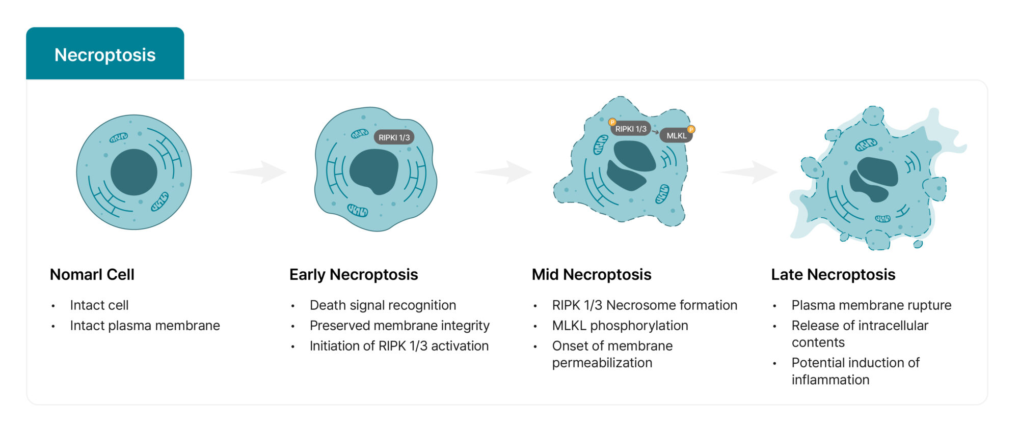
Activation of the RIPK3–MLKL axis (canonical pathway):
Depending on the cellular context, RIPK1 may function upstream, promoting RIPK3 activation and necrosome formation
MLKL (mixed lineage kinase-like protein) acts as the key executioner
Phosphorylation and oligomerization of MLKL drive plasma membrane damage, ultimately leading to lytic cell death
Early signaling and complex assembly are ATP-dependent, whereas cellular energy status may change during later stages depending on experimental conditions and time course
Major triggers
Activation of death receptors, such as TNF-α or FasL, particularly under conditions of caspase-8 inhibition
Viral infection, in which necroptosis can serve as a backup mechanism when apoptosis is evaded
Ischemia–reperfusion injury
Inflammatory response
Like necrosis, necroptosis can progress to a lytic end stage characterized by plasma membrane rupture and release of intracellular contents, which in turn promotes inflammatory responses in surrounding tissues. In this regard, necroptosis stands in contrast to the relatively “silent” nature of apoptosis and plays a particularly important pathological role in the contexts of infection, immune-mediated diseases, and ischemia–reperfusion injury.
Figure 4. Stage-dependent morphological changes during necrosis
4. Comprehensive Experimental Comparison of the Cell Death “Big Three” (Assays for Distinction)
To reliably distinguish apoptosis, necrosis, and necroptosis in the laboratory, it is safest to combine multiple lines of evidence:
(1) phenotypic features, such as plasma membrane integrity and phosphatidylserine (PS) exposure;
(2) pathway-specific molecular markers; and
(3) functional validation, including the use of inhibitors or genetic approaches.
Relying on a single assay to make a definitive conclusion is generally discouraged.
A. Basic approach: Annexin V / PI double staining
Annexin V / propidium iodide (PI) double staining is a standard method used in flow cytometry or image-based cytometry to simultaneously assess changes in plasma membrane integrity and PS exposure.
Interpretation of staining patterns
Annexin V− / PI−: Viable (live) cells
Annexin V+ / PI−: Early apoptosis (PS externalization with preserved membrane integrity)
Annexin V− / PI+: Primary necrosis-like or membrane-compromised populations
(Depending on experimental conditions, time course, and staining protocols, PS exposure may be weak or variable; additional confirmation is recommended.)
Annexin V+ / PI+: Late-stage apoptosis, secondary necrosis, or regulated lytic cell death (including necroptosis)
In Annexin V+ / PI+ populations, Annexin V binding may reflect post-rupture access to internal PS, rather than genuine early apoptotic signaling. Therefore, classifying Annexin V+ / PI+ cells as apoptotic based on this assay alone can be misleading.
Key point: Annexin V / PI staining provides a highly useful “snapshot” classification, but Annexin V+ / PI+ populations should always be interpreted in conjunction with additional analyses.
B. Pathway-specific validation: Western blotting and inhibitor assays
Western blotting and inhibitor-based assays are useful for distinguishing apoptosis from necroptosis. However, because chemical inhibitors are often context-dependent and may exhibit off-target effects, these approaches are best used to support or exclude pathway involvement, rather than to make definitive claims.
1) Caspase assay
Measurement of caspase-3/7 activity is used to assess the likelihood of caspase-dependent apoptosis.
2) Inhibitor assay
If cell death is reduced following treatment with Z-VAD-FMK (a pan-caspase inhibitor), this suggests an increased likelihood of caspase-dependent cell death.
If cell death is reduced following treatment with necrostatin-1 (a RIPK1 inhibitor), this suggests involvement of RIPK1-dependent regulated lytic cell death, including necroptosis.
Whenever possible, these results should be cross-validated using Western blot markers.
Necroptosis markers: Detection or increased levels of phosphorylated RIPK3 (p-RIPK3) and phosphorylated MLKL (p-MLKL)
Because phosphorylation sites may vary depending on species and antibody specificity, it is important to use validated antibodies appropriate for the experimental model (e.g., human or mouse). When possible, assessing total RIPK3 and total MLKL alongside phosphorylated forms is also recommended to facilitate relative comparisons.
Conclusion and research implications
Accurate identification of cell death pathways plays a critical role in interpreting drug mechanisms of action (MOA) and in studying disease pathophysiology. Although apoptosis, necrosis, and necroptosis all ultimately result in cell death, they differ substantially in the timing of plasma membrane disruption, molecular signaling cascades, and inflammatory outcomes, all of which can strongly influence experimental interpretation.
Moreover, cell death markers often change over time—for example, apoptosis may progress to late-stage apoptosis or secondary necrosis—and necrosis and necroptosis frequently exhibit similar morphological features, making them difficult to distinguish based on a single observation alone. Therefore, combining phenotypic assays such as Annexin V / PI staining with pathway-specific markers (e.g., caspase cleavage or p-MLKL) and functional validation approaches, including inhibitors or genetic perturbations, provides a more reliable framework for drawing robust conclusions.
Looking for an automated cell counter for accurate cell viability assessment?
Discover a board portfolio of cell counters and cell analysis solutions at www.logosbio.com.
REFERENCES
1) Ferroptosis, Necroptosis, and Pyroptosis in Gastrointestinal Cancers: The Chief Culprits of Tumor Progression and Drug Resistance(2023), DOI: 10.1002/advs.202300824
2) H Zhao, T Jaffer, S Eguchi, Z wang, et al. Role of necroptosis in the pathogenesis of solid organ injury. Cell Death and Disease (2015) 6, e1975; doi:10.1038/cddis.2015.316
3) Mechanisms of Cell Death: Necrosis & Necroptosis | CST Blog
4) Yuan-Shen Chen, et al. Pan-Caspase Inhibitor zVAD Induces Necroptotic and Autophagic Cell Death in TLR3/4-Stimulated Macrophages, Molecules and Cells, Volume 45, Issue 4, 2022, Pages 257-272, ISSN 1016-8478,doi.org/10.14348/molcells.2021.0193
5) Hoblos, Hanadi et al. Protein shapeshifting in necroptotic cell death signaling, Trends in Biochemical Sciences, Volume 50, Issue 2, 92 – 105
6) Kunhou Yao et al. RIPK1 in necroptosis and recent progress in related pharmaceutics, Front. Immunol., 11 February 2025, Sec. Cancer Immunity and Immunotherapy, Volume 16 – 2025 | https://doi.org/10.3389/fimmu.2025.1480027
7) Salah Abdalrazak Alshehade et al. Flow cytometry-based quantitative analysis of cellular protein expression in apoptosis subpopulations: A protocol, Heliyon, Volume 10, Issue 13, 2024, e33665, ISSN 2405-8440, https://doi.org/10.1016/j.heliyon.2024.e33665.