Choosing the right viability stain for accuracy, automation, and modern cell counting workflows
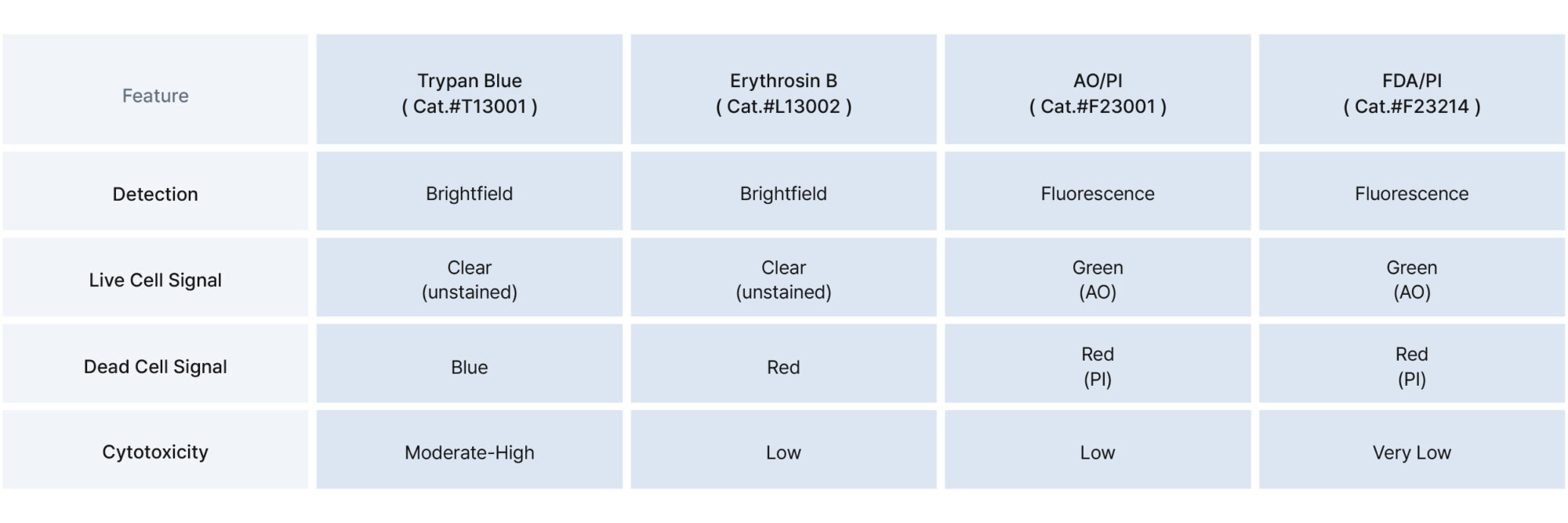
Accurate cell viability measurement is a critical part of most cell-based workflows—from routine maintenance to drug discovery and cell therapy manufacturing. For decades, Trypan Blue (TB, Cat.#T13001) has been a reliable method for distinguishing live from dead cells based on membrane integrity. It remains a widely used, accessible stain that works well with manual cell counting. It is particularly suitable for simple, short-term assays using robust cell types, where rapid manual assessment is required and fluorescence equipment is unavailable.
However, as biological assays become more complex and instrument-dependent,Trypan Blue‘s limitations have become increasingly apparent. It is less appropriate for sensitive primary or stem cells, long assay durations, or any workflow that relies on image-based or fluorescence-based automated cell counting. Issues like cytotoxicity, short counting windows, and incompatibility with fluorescence imaging make it less ideal for modern cell counting labs.
In response, alternative stains like Erythrosin B (EB, Cat.#L13002) and fluorescence-based solutions such as AO/PI (Cat.#F23001) and FDA/PI (Cat.#F23214) have emerged. These newer methods provide greater stability, lower toxicity, and compatibility with automated cell counting and fluorescence platforms.
Trypan Blue (TB, Cat.#T13001)is a diazo dye that selectively penetrates dead cells, staining them blue while live cells exclude the dye. It’s widely used for quick viability checks, particularly with hemocytometers in manual cell counting. It is best suited for simple, short-duration experiments where immediate observation is possible, and is often adequate for robust and immortalized cell lines such as HeLa or CHO cells, which are more tolerant to minor stress.
However, TB becomes problematic in cases involving delicate primary cells, stem cells, or when counting must be delayed—such as during batch processing or imaging queue workflows. In these contexts, TB‘s cytotoxicity and time sensitivity can introduce significant variability or underestimation of viability.
As shown in the graph below, PBMCs stained with TB exhibit approximately a 7 % reduction in total cell count within just 15 minutes, and close to a 20 % drop by 30 minutes. This highlights the dye’s cytotoxicity and time-sensitive behavior, which can compromise data reliability in viability-based cell counting workflows.
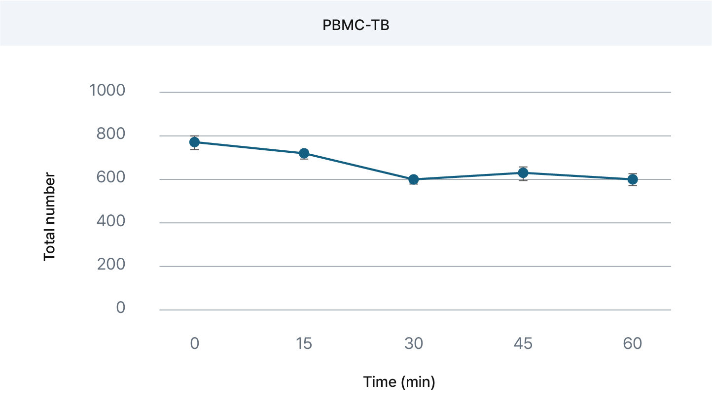
PBMCs + TB : ~7 % decrease at 15 min., ~20 % at 30 min. – sensitivity signal degradation observed.
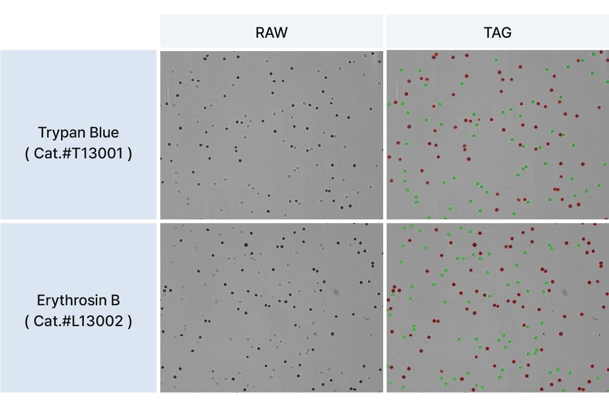
Erythrosine B (EB, Cat.#L13002) is a red dye that functions similarly to TB but with much lower toxicity. It offers a longer readout window and works well with automated image-based cell counting systems. The red color of EB may offer improved visual contrast in some brightfield imaging systems, potentially aiding cell segmentation and recognition in automated workflows.
EB is especially useful in applications where immediate counting is not possible. For example, when processing multiple samples or using shared imaging instruments, EB is reported to have greater staining stability than TB, which may help maintain data consistency in workflows that involve imaging delays. Furthermore,EB is better suited for sensitive cell types—such as PBMCs, iPSCs, and other primary cells—that are prone to TB-induced damage.
• Low cytotoxicity
• Reported stain stability of up to 30 minutes
• Red coloration may enhance detection in brightfield imaging
• Compatible with bright-field automated cell counting platforms
EB provides comparable image quality to TB in grayscale brightfield imaging and supports reliable cell classification in automated analysis workflows.
Acridine Orange (AO, Cat.#F23002) stains all cells, fluorescing green in viable cells, while Propidium Iodide (PI, Cat.#F23003) only enters dead cells and fluoresces red. AO/PI enables simultaneous live/dead identification in fluorescence-based cell counting. Because it uses two clearly distinguishable channels, AO/PI staining is especially useful in high-throughput screening and kinetic assays where tracking viability over time is essential.
This method is widely adopted in both research and industry settings for its robustness and compatibility with flow cytometry, fluorescence microscopes, and image-based automated counters. It is also considered a preferred choice for samples with mixed viability states or when greater resolution is required between live and apoptotic populations.
If you’re looking for a deeper dive into how AO/PI
staining works at the molecular level, including protocols and best practices, refer to our detailed guide:
” How to Measure Cell Viability Using Acridine Orange/Propidium Iodide: The Principle and the Procedure. ”
• High sensitivity and contrast
• Real-time dual-channel viability analysis
• Ideal for flow cytometry and high-content cell counting systems
As shown in the graph below, PBMCs stained with AO/PI
maintain a highly stable total cell count across 30 minutes incubation. This indicates minimal cytotoxicity and excellent temporal consistency, making AO/PI
a reliable choice for fluorescence-based viability assessment in cell counting workflows.
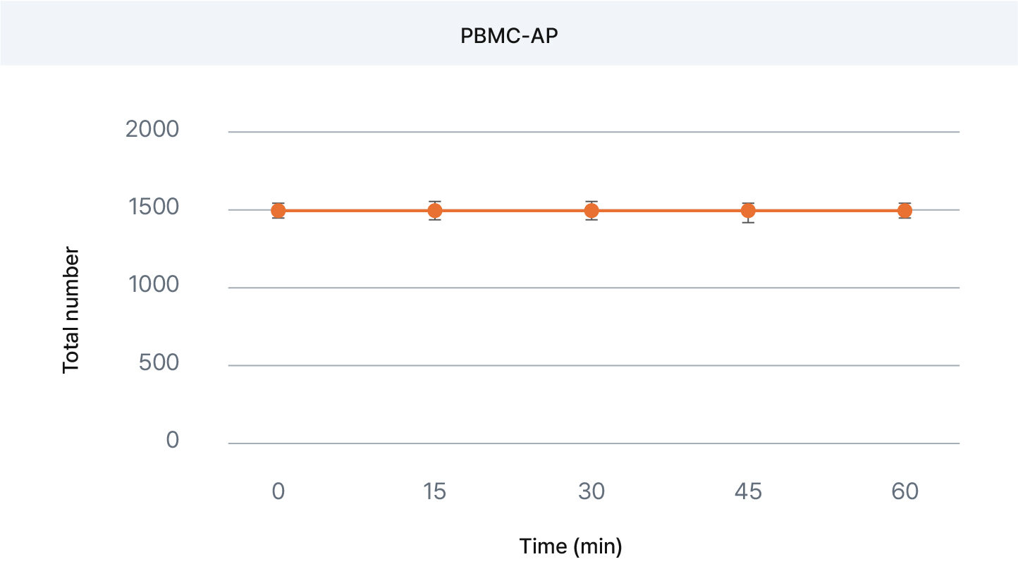
PBMCs + AO/PI : Stable total count over 30 min. No significant viability shift observed.
Fluorescein Diacetate (FDA) is a non-fluorescent compound converted into fluorescein (green) by intracellular esterases in live cells. PI stains dead cells red, creating a clear viability map ideal for cell counting automation.
Unlike AO, which binds directly to nucleic acids, FDA offers a readout that is dependent on esterase activity, meaning only metabolically active live cells are labeled. This makes FDA/PI an excellent choice for experiments requiring a high degree of specificity for cell health.
FDA/PI (Cat.#F23214)is particularly useful in scenarios where background staining must be minimized and when working with small or low-signal cells such as yeast. In fact, this staining method is the basis of the Logos Biosystems Yeast Viability Kit 1 (Cat.#F23202), which is specifically optimized for high-contrast fluorescence-based cell counting of yeast cells.
For detailed application data, see our Application Note :
“ Fast Cell Counting and Viability Measurement of Yeast Cells with the LUNA-FL™ Fluorescence Cell Counter ”
• Very low toxicity
• Enzyme-specific live cell signal
• Long staining window
• Highly compatible with automated cell counting platforms
Trypan Blue (Cat.#T13001) remains a familiar and accessible stain, and it still plays a useful role in simple, manual cell counting workflows—especially for robust cell lines and short-term assays. However, it falls short when used with sensitive primary cells, delayed imaging conditions, or high-throughput demands.
For researchers seeking improved accuracy, lower cytotoxicity, and compatibility with automation, alternative stains offer meaningful advantages.
• Erythrosin B (Cat.#L13002) improves safety and stability without requiring fluorescence, making it a practical next step for labs transitioning from manual to image-based cell counting.
• On the other hand, AO/PI
(Cat.#F23001) and FDA/PI (Cat.#F23214) deliver high-contrast, fluorescence-based solutions suitable for advanced analytical platforms, including flow cytometry and automated imaging systems.
Selecting the right viability stain isn’t just about tradition—it’s about ensuring your cell counting method aligns with your sample type, instrumentation, and experimental goals.
For additional guidance on viability assay selection and broader context, see our comprehensive guide:
” The Ultimate Guide to Cell Viability Measurement. ”
Q1: Can I use Trypan Blue with fragile cells like PBMCs or iPSCs?
A: It is not recommended, as TB can be cytotoxic.
EB or AO/PI
offer better viability preservation.
Q2: Is FDA/PI suitable for all cell types?
A: FDA/PI is particularly effective for yeast and small cells with low background fluorescence. For mammalian cells AO/PI is generally preferred.
Q3: How do I choose between EB and AO/PI?
A: Choose EB if you are working with a brightfield counter and want to avoid fluorescence. Choose AO/PI for fluorescence-based detection and more detailed viability profiling.
Looking for the Korean version?Click here.
Logos Biosystems provides a diverse portfolio of automated cell counters designed to meet various laboratory requirements.
To learn more, visit www.logosbio.com.


